bone tumors in cats and dogs
What bone tumors occur in dogs and cats?
Tumors (neoplasms) of the bone can be divided into two categories: primary bone tumors, which originate within a bone, and secondary (metastatic) bone tumors, which are the result of the spread of a cancer that has originated elsewhere in the body. Primary bone tumors may be benign or malignant, while all secondary tumors are malignant.
In dogs, over 80% of primary bone tumors are osteosarcomas, an aggressive cancer of bone-producing cells. These occur most frequently in middle-aged to older large and giant breed dogs, and tend to develop just above the wrist joint, just below the shoulder joint, and adjacent to the knee (stifle), although they can arise in any bone, including the bones of the skull, ribs, and vertebrae. Osteosarcomas usually grow rapidly and are at a high risk of metastasizing, most commonly to the lungs. The second most common type of primary bone tumor in dogs is the chondrosarcoma. This malignant tumor of cartilage-producing cells accounts for 5-10% of primary bone tumors in dogs. These tumors are most frequently encountered in middle-aged to older dogs. While chondrosarcomas most commonly develop in the nasal cavity, they can arise anywhere in the body where cartilage is present, including the joints and ribcage. Chondrosarcomas tend to be slower to metastasize than osteosarcomas. Other, less common types of primary bone tumors include hemangiosarcoma (a cancer of blood vessel walls), fibrosarcoma (a cancer of connective tissue), multilobulated osteochondrosarcoma (a cancer of the periosteal lining of a bone), and cancers of circulating immune cells (lymphoma, plasma cell tumors).
In cats, 70-80% of primary bone tumors are osteosarcomas. These occur most frequently in middle-aged cats, and tend to arise adjacent to the knee, in the upper arm, or in the skull. While locally invasive, osteosarcomas are much less likely to metastasize in cats than they are in dogs. The second most common type of primary bone tumor in cats is the fibrosarcoma, arising in connective-tissue producing cells. Chondrosarcomas and hemangiosarcomas can also arise within bone, but are rare.
What are the signs of a bone tumor?
The most common signs of a bone tumor include lameness, swelling, and pain at the site of tumor growth. This typically develops gradually over time, although a sudden worsening of lameness may occur if a pathological fracture occurs through the abnormal, weakened bone.
How are bone tumors diagnosed?
The first step in diagnosing a suspected bone tumor is to take x-rays of the area. On x-rays, a bone tumor often appears as an area of abnormal darkening within the normally bright bone, representing the breakdown (lysis) of normal bone. These areas of lysis may have smooth contours or may appear mottled and irregular. New bone formation may also be present, extending outward from the exterior surface of the bone. In some cases, a fracture is present through the abnormal area due to weakening of the bone structure.
When a suspected bone tumor is identified on x-rays, the next step is to obtain a biopsy sample from the lesion. Biopsy collection requires that the patient be briefly placed under general anesthesia while a small incision is made over the area. Tissue samples are collected either with a very large biopsy needle or using a scalpel.
If the biopsy report indicates the presence of a malignant growth, additional testing, such as chest x-rays and abdominal ultrasound, are preformed to look for evidence of metastases to aid in treatment planning.
How are bone tumors treated?
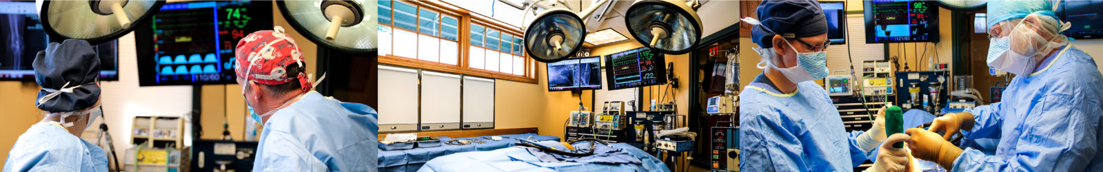
The mainstay of treatment for bone tumors is the complete surgical removal of the tumor. Because most bone tumors develop on a limb, this usually requires amputation of the affected limb. If the tumor is on a toe bone, amputation of the toe may be all that is required. For tumors of the ribs, vertebrae, skull, and jaw, advanced imaging, such as a CT scan or MRI, is necessary to determine the precise dimensions and full extent of the tumor. This will enable the surgeon to determine whether removal is possible and plan the procedure. If a tumor is deemed non-resectable, radiation therapy is an alternative option that may shrink or destroy the tumor; the availability of radiation therapy in veterinary medicine is limited to large urban areas and universities, but if travel is an option, this can be an effective treatment modality.
Depending on the type of tumor, additional treatment may be recommended following surgery. Because of the high risk of metastasis with osteosarcoma and hemangiosarcoma, chemotherapy is typically recommended once the surgery site is healed. This involves a series of intravenous injections of chemotherapy drugs about every two or three weeks for four to six treatments. The side effects of chemotherapy are usually milder in animals than they are in people, and are typically limited to several days of inappetance or GI upset, which is usually controllable with antinausea medication. If moderate to severe side effects do develop, future doses of chemotherapy are reduced to avoid recurrence. Chemotherapy also affects the bone marrow, causing a temporary decrease in the production of white blood cells, which protect the body from infection. A complete blood count is monitored following each dose of chemotherapy, and if the white blood cell count becomes too low, the patient is placed on a protective antibiotic and future doses of chemotherapy are decreased to reduce the risk of recurrence. Because chemotherapy agents are toxic to healthy cells in both animals and humans, special precautions will be necessary in handling a pet who has recently received chemotherapy. Your veterinarian will review these measures with you prior to starting chemotherapy.
What is the prognosis for bone tumors?
The prognosis for a bone tumor depends on its identity. For tumors that either do not metastasize or are slow to do so, such as chondrosarcomas, multilobular osteochondrosarcomas, and osteosarcomas in cats, following complete removal, the prognosis is very good to excellent. Chest X-rays are typically recommended every 4-6 months to check for the appearance of any metastases, but most of these pets do not develop further disease in the future.
In dogs, osteosarcomas have a much more guarded prognosis, due to their tendency to spread to other parts of the body early in their growth. With surgery alone, the average survival time for osteosarcoma is 4 months. With surgery and chemotherapy, average survival is extended to 9-12 months. In 5-10% of osteosarcoma cases, the cancer has not yet microscopically spread at the time of amputation; these patients often go on to live for years without recurrence of the cancer.
Hemangiosarcomas also tend to metastasize early in the course of disease. With surgery and chemotherapy, average survival time is 6-9 months.

Let our highly trained and experienced team of veterinarians and veterinary technicians help you keep your cat as happy and healthy as they can be.
Call the Animal Clinic of Billings and Animal Surgery Clinic to schedule your pet cat’s next wellness examination with one of our veterinarians today!
406-252-9499 REQUEST AN APPOINTMENT
ANIMAL CLINIC OF BILLINGS AND ANIMAL SURGERY CLINIC
providing our region’s companion animals and their families what they need and deserve since 1981
1414 10th St. West, Billings MT 59102
406-252-9499