Internal Fixation Surgery
What is Internal Fixation for Fractures?
Internal fixation is a strategy for fracture repair that involves surgically accessing the fracture site to realign the bone segments (called “open reduction”) and rigidly stabilizing them with surgical implants to enable bone healing to occur. This is in contrast to closed reduction with external fixation, which does not involve surgically accessing and realigning the break, but rather relies on an external fixator (composed of bone pins fixed to metal rods located external to the skin) or cast applied to the limb to immobilize the bone fragments during the healing process.
The best approach to the treatment of a specific fracture depends on many variables, including:
- Whether the fracture is complete (all the way through the bone) or incomplete (only part of the way through the bone)
- Whether the fracture segments are displaced (out of normal anatomic alignment) or nondisplaced (in normal alignment, with the fractured surfaces in contact with each other)
- Whether the fracture is open (has penetrated through the skin, also called “compound”) or closed (has not penetrated through the skin)
- The location, orientation, and number of fractured segments
- Whether the fracture segments are reducible (can be put back together to enable weight bearing while healing) or non-reducible (cannot be reconstructed into a solid configuration, necessitating surgical implants to completely take over load bearing throughout the healing process)
- Whether the fracture involves a joint surface, active growth plate (physis), or tendon/ligament attachment site
- The degree of damage to the soft tissues and blood supply in the area of the fracture
- Whether the patient has other orthopedic or neurologic conditions
- The patient’s age, body mass, and general health
As mentioned above, fracture management can be divided into two categories: closed reduction with external fixation and open reduction with internal fixation.
In closed reduction with external fixation, either external coaptation or an external fixator is used for stabilization until the fracture heals. External coaptation involves placing either a cast or splint on the affected limb to hold the fracture site as still as possible and protect it from external forces that could disrupt healing. External fixators are composed of a system of surgical pins that extend through the bone fragments (perpendicular to the long axis of the bone) and out through the skin, where they are rigidly attached to metal bars running parallel to the limb, which hold them securely in order to stabilize the bone fragments in the correct positions for healing and to take over the forces of weight bearing while the fracture site heals.
Fractures that can usually be managed successfully with closed reduction include incomplete fractures and some nondisplaced fractures that occur below the elbow and knee (stifle). External fixation is also sometimes used for non-reconstructible, severely comminuted fractures (composed of many small pieces), with the goal of maintaining the length and alignment of the limb while taking over load-bearing duties, giving the body time to fill in the site with new bone.
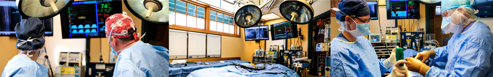
Most fractures in animals require open reduction with internal fixation, which involves surgically accessing the fracture site to reconstruct the major fragments and secure them in place with surgical implants. Surgical fracture stabilization is required more frequently in animals than in humans because of the impossibility of preventing limb use during the healing process. A number of different methods of rigid internal fixation exist, and the best ones to use in a specific case will depend on the specific configuration of the fracture. The most commonly-used fixation devices include intramedullary pins (metal rods that can be driven down the marrow cavity), cerclage wires (wires that are looped around the shaft of a long bone and twisted tight to hold highly oblique fragments together), metal plates, interlocking nails (metal rods extending the length of the marrow cavity with interlocking cross-screws at either end), bone screws, and pins.
Following a physical exam and sedated x-rays of the area, the surgeon will be able to determine the best treatment plan, taking into account all of the variables discussed above. Using bone models and x-rays, they will be able to walk you through the procedure so you understand exactly what they will be doing to fix your pet’s fracture.
What is the recovery like following open reduction with internal fixation?
Most pets are able to go home the day after surgery unless they have other injuries or problems that require further hospitalized care. Most limb fractures will be bandaged after surgery to limit swelling and protect the skin incision from contamination and interference by the patient. Fractures involving the humerus (upper arm bone), scapula (shoulder blade), femur (thigh bone), and pelvis are typically not bandaged due to practical limitations in maintaining bandages over these locations. All patients are sent home with pain medications to ensure they remain comfortable throughout the post-operative period. If deemed necessary, they may also be sent home on an antibiotic. The bandage will be removed after about five to seven days, and the sutures will be removed at the patient’s two week recheck.
Following fracture stabilization, strict activity restriction is essential to a successful outcome. Any method of fracture fixation—whether a cast, external fixator, or bone plate—will fail if a patient is too active and exerts too much force on the area. For the first four weeks after the fracture is stabilized, only short, slow leash walks should be permitted in order to use the bathroom. At all other times, the pet should be confined to a crate or small room that does not contain anything they could jump up on. It can be a real challenge to keep a pet confined for an extended period of time like this, but it is absolutely essential. Sometimes, a sedative or anti-anxiety medication can be prescribed to help keep the pet calm and relaxed during their convalescence. Passive stretches and range of motion exercises may also be incorporated, depending on the location of the fracture.
After four weeks, a set of x-rays will be taken to evaluate the condition of the implants and the progression of bone healing. If fracture healing is progressing well, you will likely be instructed at this point to start increasing the length of leash walks, although it will still be important to avoid any running, jumping, or playing with other pets. Physical rehabilitation exercises, including sessions in an underwater treadmill, may be added to start rebuilding lost muscle mass and encourage controlled limb use.
X-rays will be repeated at eight to ten weeks post-repair, and further increases in physical activity will be permitted based on the progress of bone healing. Often, puppies and kittens will be completely healed at this point, and can return to full activity, while healing in mature pets typically takes 12-16 weeks. Monthly x-rays will be repeated until the fracture is fully healed.
What are the potential risks and complications of open reduction with internal fixation?
Complications that may occur following open reduction with internal fixation include infection, non-union, delayed union, malunion, implant failure (loosening or breakage), premature growth plate (physis) closure, and arthritis.
Infections associated with fractures may be limited to the skin at the site of a wound or surgical incision or may infect the deeper tissues and the bone itself. Conditions that increase the risk of infection include open fractures (where the bone has penetrated the skin and been exposed to the external environment) and the presence of significant damage to the soft tissues and blood supply around the fracture. Bacterial infections involving bone require an extended course of antibiotics—often 4 weeks or more. If a fracture site containing surgical implants becomes infected, implant removal may become necessary to fully eliminate the infection.
A delayed union is a fracture that heals more slowly than expected. Factors that can contribute to a delayed union include concurrent illness, advanced age, inadequate nutrition, extensive damage to the soft tissues and blood supply of the fracture area, loose or unstable surgical implants, and too much physical activity. Some of these factors can be addressed in order to accelerate the rate of healing. Inserting a bone graft at the fracture site can also help to facilitate bone growth. Given enough time, a delayed union will eventually heal, but it may take months longer than expected.
A non-union occurs when the healing process comes to a halt before the fracture segments have fused back together. This is diagnosed when no progress is observed on consecutive sets of monthly x-rays. Potential causes of a non-union include instability of the fracture segments and insufficient blood supply to the fracture site. Instability may result from the use of implants that are not strong enough or that are not ideally suited to the configuration of the fracture, as well as from too much activity causing excessive stress on the implants. Insufficient blood supply to the fracture site is typically a consequence of the trauma that caused the fracture. This is also thought to play a role in the disproportionately high incidence of non-unions in forearm fractures of toy and small breed dogs; it is thought that circulation to this area is generally less robust in these breeds than in larger dogs. Non-unions require surgical intervention in order to heal. If instability is the cause of a non-union, the implants are removed, any fibrous tissue growing in and around the fracture site is excised, and stronger surgical implants are applied. If an insufficient blood supply is to blame, surgery is performed to remove fibrous tissue and open up the marrow cavities at the fracture site. If necessary, new surgical implants may be placed to hold the fracture ends in close contact. A cancellous bone graft, consisting of low-density “spongy” bone found within the ends of many bones in the body, is harvested from the top of the humerus or the pelvis, and placed in the fracture site to provide active bone-forming cells and growth factors to jump-start the healing process.
A malunion occurs when the fracture heals in improper alignment. This can happen if bending, breakage, or loosening of implants occurs (often due to overuse of the limb) and is not detected before healing has finished. A malunion may cause functional impairment of the limb and arthritis in adjacent joints. Malunions are treated with a surgical procedure called a corrective osteotomy, in which the bone is cut and re-set in proper alignment. Surgical implants are used to stabilize the osteotomy site while it heals.
Implant failure occurs when a metal surgical implant (pin, plate, screw, or wire) either loosens, bends, or breaks before the fracture has fully healed. This can happen if an improper type or size of implant is used, or if the patient is too active and exerts excessive strain on the repair site. This typically results in instability at the fracture site and sometimes even displacement of the fracture segments. If implant failure occurs, a second surgery is needed to realign the fracture and replace the damaged implants.
Premature growth plate closure can occur if a fracture runs through an active growth plate in a growing puppy or kitten. Growth plates are the centers of bone growth found toward either end of a long bone that are responsible for the increase in bone length that occurs as an animals grows. In some cases, trauma to these areas can cause them to shut down too early, resulting in the bone not growing to its full adult length. If this occurs in a non-paired bone, such as the humerus (upper arm bone) or femur (thigh bone), the affected limb will end up shorter than the opposite limb. If premature growth plate closure occurs in one of the paired bones (the radius or ulna of the forearm, or the tibia or fibula of the lower leg), it results in bowing of the limb as the unaffected bone continues to grow. The severity of this limb shortening or bowing will depend on how close the bone is to maturity when the injury occurs. If impaired limb use results, options exist for surgical correction of the abnormality.
Any fracture that runs through a joint surface will cause arthritis to develop over time. With perfect surgical realignment, this arthritis is often minimal, and easily managed with anti-inflammatory medications and joint health supplements. If fracture alignment at the joint surface is slightly off, much more severe arthritis can develop that results in pain and impaired limb use.
What is the prognosis following open reduction with internal fixation?
The prognosis following open reduction with internal fixation for fracture repair is very good to excellent for simple fractures in healthy animals and good for complex fractures, fractures involving a joint, and fractures in older or pets and those with chronic health problems. The most important factor in achieving a successful outcome after surgery is strict activity restriction until x-rays show advanced bone healing. Most pets return to completely normal function within three to four months. Fractures that involve a joint will result in the development of varying degrees of arthritis. With perfect or near-perfect alignment, which is achievable only with open reduction and internal fixation, arthritis will be minimal and will be controllable with anti-inflammatory medications and joint health supplements.
Let our highly trained and experienced team of veterinarians and veterinary technicians help you keep your cat as happy and healthy as they can be.
Call the Animal Clinic of Billings and Animal Surgery Clinic to schedule your pet cat’s next wellness examination with one of our veterinarians today!
406-252-9499 REQUEST AN APPOINTMENT