lumbosacral stenosis and spine disease (LS)
What is lumbosacral stenosis?
Lumbosacral stenosis, also known as cauda equina syndrome, is a narrowing of the spinal canal where the lumbar spine (low back) meets the sacrum (tailbone) at the top of the pelvis. Factors that frequently contribute to this narrowing include upward protrusion of the intervertebral disc, thickening of the joint capsules and ligaments connecting the lumbar vertebra to the sacrum, bone overgrowth along the periphery of these joint surfaces, and joint instability. This narrowing causes compression of the bundle of nerve roots (called the cauda equina) coursing down the spinal canal from the end of the spinal cord (at the level of the 5th or 6th vertebra). Compression of these nerve roots has the potential to cause pain, hind limb weakness, lameness, urinary and fecal incontinence, and loss of tail function.
What causes lumbosacral stenosis?
Several factors likely contribute to the development of lumbosacral stenosis. Intervertebral disc protrusion is believed to be caused by a combination of age-related dehydration of the central disc region as well as thickening of the peripheral disc structure from years of mechanical stress. The thickening of the joint capsules and ligaments surrounding the lumbosacral junction is thought to be an attempt by the body to counteract joint instability. The bone overgrowth associated with these joints is also a consequence of joint instability and consequent arthritis.
Who is at risk of lumbosacral stenosis?
While lumbosacral stenosis can occur in any dog or cat, it is usually encountered in middle-aged to older large and giant breed dogs. The incidence is highest in the German Shepherd Dog. Other breeds that appear to be at increased risk of developing this condition include Labrador Retrievers, Golden Retrievers, Great Danes, Boxers, Irish Setter, Airedale Terrier, and English Springer Spaniel.
What are the clinical signs of lumbosacral stenosis?
Lumbosacral stenosis typically causes a gradual onset of progressive hind limb lameness and weakness that is worsened by physical activity. Affected dogs often have difficulty rising and climbing stairs, and display a short-strided gait in the rear. A hunched posture may be noticed as an affected individual flexes their spine in an attempt to alleviate the compression. A change in tail carriage and movement is not unusual. As the condition progresses, urinary and/or fecal incontinence may develop.
How is lumbosacral stenosis diagnosed?
The first step in diagnosing lumbosacral stenosis is a thorough physical exam. Affected patients demonstrate pain on palpation of the lumbosacral junction, both from above and from below (on rectal exam), and on raising of the tail up over the hindquarters. Neurologic deficits may be present in the hind limbs, and a decrease in muscle mass is often noted. On a gait exam, hind limb weakness and an abnormally short stride are frequently observed. The toenails on a patient’s rear paws may be worn short from scuffing their paws as they walk.
If a patient’s physical exam is consistent with lumbosacral stenosis, the next step is to take precisely-positioned x-rays of the low back and pelvis. Sedation is typically required due to patient discomfort. While x-rays cannot provide a definitive diagnosis of lumbosacral stenosis, they are useful to rule out other possible causes of a patient’s signs, such as a fractures, infections, or tumors of the vertebrae. X-ray findings that are frequently encountered with lumbosacral stenosis include mineralization of intervertebral disc material, narrowing of the lumbosacral disc space, increased density (sclerosis) of the vertebral endplates (where the vertebral bodies meet the intervertebral disc), spondylosis (new bone formation) extending from the vertebral endplates, and a “stair-step” misalignment of the spinal canal.
The definitive diagnosis of lumbosacral stenosis requires either a CT scan or an MRI, which provides a detailed view of both the bones and soft tissue structures within and around the lumbosacral junction. These scans require heavy sedation or anesthesia, as the patient must remain perfectly still throughout the image acquisition period.
How is lumbosacral stenosis treated?
Treatment for lumbosacral stenosis may be either surgical or non-surgical. In patients who do not have any neurologic deficits, conservative management is typically instituted. This involves medications for pain and inflammation, a diet for patients who are overweight, and strict activity restriction for at least six to eight weeks. In some patients, periodic epidural steroid injections are also administered. Conservative management is successful in up to 50% of patients.
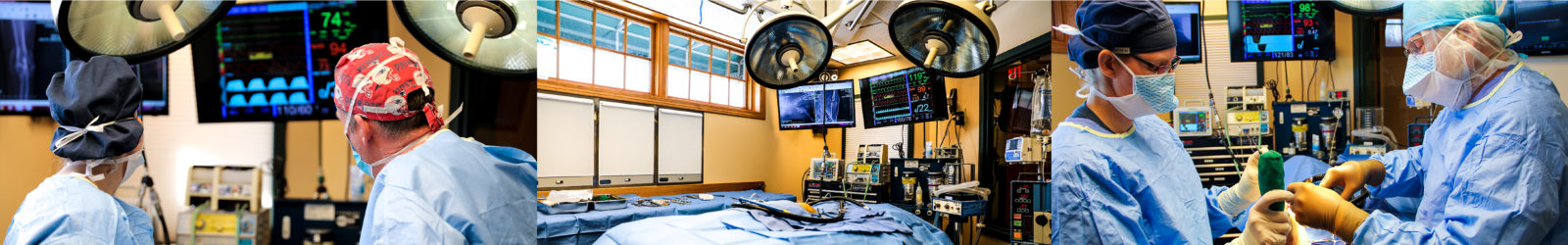
Patients who have neurologic deficits, as well as those for whom conservative treatment has been ineffective, may benefit from surgery to relieve the compression. Several different surgical techniques can be utilized, and the most appropriate ones for a specific patient will depend on the factors contributing to the compression and the judgement of the surgeon. If there is protrusion of the lumbosacral disc, a procedure called a dorsal laminectomy is most commonly performed. This involves removing the bone forming the roof of the spinal canal at this location to give the nerves more space. A dorsal annulectomy, in which the top portion of the bulging intervertebral disc body (pushing up on the nerves from below) is carefully removed, may also be performed to further alleviate compression. In order to address the vertebral joint instability causing the progressive overgrowth of the bone and soft tissues, a distraction and stabilization procedure is performed at the lumbosacral junction. This involves the use of screws, pins, wire, and/or bone cement to rigidly stabilize the final lumbar vertebra and the sacrum together in a neutral position. Cartilage is removed from the joint surfaces, and bone graft is placed to promote permanent fusion of these two structures. Additional procedures may be performed as needed to release nerve roots and spinal nerves that have been entrapped by overgrown soft tissues and bone.
What post-operative care is required?
Most patients are able to go home 24 to 48 hours following surgery. They will be sent home with pain medication, an anti-inflammatory, and an antibiotic. The most important aspect of post-operative care is strict activity restriction for eight weeks. This is important to minimize the risk of implant breakage while allowing time for bony fusion to occur. X-rays will be taken at four, eight, and sometimes twelve weeks post-surgery to assess bone fusion and implant integrity. Initially, only short, slow leash walks will be permitted. Following rechecks at two, four, and eight weeks, the amount and types of physical activity permitted will be gradually expanded. For optimal results, appointments with our physical rehabilitation department will be strongly recommended once or twice weekly to aid in healing, comfort, and reconditioning. At-home physical rehabilitation exercises to be performed between appointments will be taught to enhance each patient’s recovery.
What are the potential risks and complications of surgery for lumbosacral stenosis?
The surgery performed to address lumbosacral stenosis is very delicate, and involves manipulations in close proximity to a number of large nerves and blood vessels. Potential complications include damage to nerves and nerve roots, new or worsening compression due to implant installation and/or scar tissue formation, hemorrhage, infection, implant breakage or loosening, and failure of bone fusion. While these complications are rare, they can be potentially serious, and may necessitate a second surgical procedure.
What is the prognosis for lumbosacral stenosis?
The prognosis for lumbosacral stenosis depends on the duration and severity of their symptoms. Patients without neurological deficits who have only been affected for a few months or less have a 50% chance of recovering with conservative management. Those patients with minimal to no neurologic deficits who fail conservative management have a 70-80% chance of improvement with surgery. Patients with severe neurologic abnormalities, urinary incontinence, and/or fecal incontinence, have a much more guarded prognosis, and typically do not recover bladder and/or bowel control, although surgery may still be effective in improving comfort.
Let our highly trained and experienced team of veterinarians and veterinary technicians help you keep your cat as happy and healthy as they can be.
Call the Animal Clinic of Billings and Animal Surgery Clinic to schedule your pet cat’s next wellness examination with one of our veterinarians today!
406-252-9499 REQUEST AN APPOINTMENT
ANIMAL CLINIC OF BILLINGS AND ANIMAL SURGERY CLINIC
providing our region’s companion animals and their families what they need and deserve since 1981
1414 10th St. West, Billings MT 59102
406-252-9499