Medial Patella Luxation (MPL) Surgery
What is Medial Patellar Luxation surgery?
Medial patellar luxation (MPL) is a condition in which the patella (kneecap), which normally slides up and down within a groove running along the front of the femur within the knee joint as the knee is flexed and extended, pops out of its groove to the inside (towards the other back leg.) When the patella luxates, in addition to causing discomfort, it disrupts the normal function of the knee by pulling the quadriceps muscle group out of its normal alignment. This is a relatively common disorder in dogs, but is seen only rarely in cats.
Most cases of medial patellar luxation are genetic in origin and occur in toy and small breed dogs who are born with anatomic abnormalities including coxa vara (where the femoral neck is too horizontally oriented) and decreased femoral neck angle of anteversion (where the angle between the long axis of the femoral neck and the transverse axis of the knee joint is too small, resulting in mild external rotation of the knee relative to the hip). These abnormalities cause inward (medial) displacement of the quadriceps muscle group and patella. The aberrant forces generated by this displacement cause the leg bones to bow outward at the knee as the individual grows, worsening the misalignment. In addition, the lack of the normal pressure of the patella gliding in the femoral trochlear groove results in underdevelopment of this important structure.
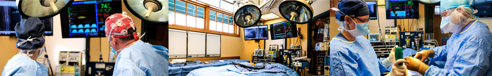
MPL surgery aims to stabilize the patella in its correct position. This is most commonly achieved using a combination of four surgical modifications:
- A trochleoplasty is performed to increase the depth of the trochlear groove. This involves the removal of a wedge or block-shaped piece of cartilage and bone from the groove, followed by deepening of the resulting defect and replacement of the original fragment. Creating a deeper groove makes it more difficult for the patella to luxate.
- A lateral tibial tuberosity transposition is performed to shift the insertion point of the patellar tendon laterally. The patellar tendon extends from the end of the quadriceps musculature to the front of the upper tibia (shin bone), and houses the patella. Its attachment point on the tibia, called the tibial tuberosity, is the bony prominence palpable just below the knee joint. In a tibial tuberosity transposition, a cut (osteotomy) is made through the bone of the tibia to detach the tibial tuberosity. The fragment is then shifted slightly laterally (toward the outside) and secured in place with surgical pins. By moving the insertion point of the patellar tendon laterally, this procedure re-aligns the patella with its groove.
- A medial desmotomy, or vertical incision through the contracted connective tissues and joint capsule on the medial (inner) side of the knee joint, releases tension that has been helping to hold the quadriceps and patella in their medially displaced position.
- A lateral imbrication, in which the stretched portion of the joint capsule on the outer (lateral) side of the knee joint is tightened up, helps to further stabilize the patella.
What patients can benefit from MPL Surgery?
Whether MPL surgery is indicated for a specific patient depends on the grade of their luxation and the magnitude of their clinical signs. Based on a physical exam, a MPL can be classified into one of four grades:
- Grade 1 – The patella can be pushed out of the groove, but immediately pops back in on its own. This can be an incidental finding in a dog without any clinical signs.
- Grade 2 – The patella pops out on its own, but returns to the groove with flexion of the knee. This is typically associated with brief and intermittent bouts of lameness, although it may become more frequent over time.
- Grade 3 – The patella is always out of the groove, but can be pushed back in. After being pushed into its groove, it immediately pops back out. This is most commonly associated with persistent lameness and discomfort.
- Grade 4 – The patella is always out of the groove and cannot be pushed back in. This usually causes persistent lameness and discomfort.
Patients with symptomatic grade one luxations and mild grade two luxations can usually be managed successfully with medications, joint health supplements, nutraceuticals, and weight management.
Patients with more severe grade two luxations, as well as all grade three and four luxations, require surgery to restore comfort and limb function.
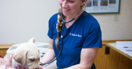
What post-operative care is required?
Following surgery, physical activity must be restricted until the bones have fully healed. X-rays are taken at 4-6 weeks and again at 8-10 weeks to monitor the healing of the tibial tuberosity osteotomy. Most dogs are able to return to full activity after their 8-10 week recheck. Until then, only low-impact on-leash exercise is allowed in order to prevent excessive pull on the tibial tuberosity as it heals. For most dogs, this means two to three short, slow walks per day. Daily range of motion exercises and stretches are incorporated to prevent muscle contracture and joint stiffness during the healing process.
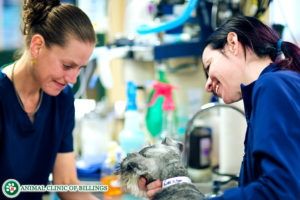
What are the risk and complications of MPL Surgery?
The most frequently encountered complication is recurrence of the luxation. With medial patellar luxations, the risk of re-luxation is about 10% for grade three luxations and is greater than 30% for grade four luxations. Other potential complications include implant loosening or breakage, tibial tuberosity avulsion (pulling away from the rest of the tibia), movement of the trochlear wedge, rupture of the patellar ligament, and progression of arthritis.
What is the prognosis after MPL Surgery?
The prognosis for comfort and a return to normal or near-normal limb use following surgery is typically very good, although arthritic changes that are already present at the time of surgery cannot be reversed, and if advanced, may contribute to ongoing discomfort. This can often be successfully alleviated using medications, supplements, physical rehabilitation therapy, and weight management.
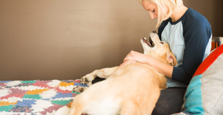
Let our highly trained and experienced team of veterinarians and veterinary technicians help you keep your cat as happy and healthy as they can be.
Call the Animal Clinic of Billings and Animal Surgery Clinic to schedule your pet cat’s next wellness examination with one of our veterinarians today!
406-252-9499 REQUEST AN APPOINTMENT
ANIMAL CLINIC OF BILLINGS AND ANIMAL SURGERY CLINIC
providing our region’s companion animals and their families what they need and deserve since 1981
1414 10th St. West, Billings MT 59102
406-252-9499