Hip Dysplasia
What is hip dysplasia?
Hip dysplasia is a common orthopedic condition in dogs in which the ball-and-socket hip joint does not form properly in a growing puppy. In these dogs, hip joint laxity, which may be caused by malformation of the hip socket (acetabulum) or ball (femoral head) or by weakness and stretching of the connective tissues of the joint capsule and hip joint ligaments, results in joint instability. The body’s response to the abnormal forces caused by this instability produces further loss of hip socket depth and flattening of the femoral head. Over time, this persistent joint instability causes the development of significant arthritis.
Most dogs diagnosed with hip dysplasia have morphologically normal hips at birth, but begin to develop abnormal hip joints within the first few months of life. Usually both hips are affected, although one can be more abnormal than the other. In most cases, symptoms become apparent by 8 months of age.
What is the cause of hip dysplasia in dogs?
Hip dysplasia is fundamentally a genetic disorder, although its severity can be influenced by external factors such as nutrition, growth rate, activity level and type, other environmental factors, and possibly neuter status. Hip dysplasia is most common in large and giant breed dogs. Breeds at particularly high risk include Labrador Retrievers, Golden Retrievers, German Shepherd Dogs, Rottweilers, Saint Bernards, Newfoundlands, Mastiff breeds, Otterhounds, American Bulldogs, and Black Russian Terriers.
An increased risk of developing hip dysplasia has been demonstrated in young dogs that have unlimited access to food and subsequently experience more rapid growth than puppies whose intake is limited. Additionally, the calcium and phosphorus requirements for large and giant breed puppies are different than for small and medium breeds, and feeding regular puppy food to these puppies may increase their risk of developing hip dysplasia. To minimize risk, it is recommended that all large and giant breed puppies be fed a puppy food specifically formulated for large and giant breed puppies, and that their intake be limited in order to maintain a lean body mass as they grow.
It appears that allowing unrestricted off-leash exercise to puppies under 3 months of age may reduce their risk of developing hip dysplasia. However, one study suggests that allowing puppies under 3 months of age to go up and down stairs may actually increase their risk. A decreased risk of developing hip dysplasia has also been reported in dogs born in the spring and summer, although the reason for this remains unknown.
Recent studies suggest that waiting to spay or neuter large and giant breed dogs until they are fully grown may also reduce their risk of developing orthopedic problems including hip dysplasia, as reproductive hormones impact skeletal growth and development.
Responsible breeders have a few tools available to them determine if a dog they want to breed is likely to produce puppies that will be affected by hip dysplasia. The Orthopedic Foundation of America (OFA) and the Pennsylvania Hip Improvement Program (PennHIP) both make assessments of hip conformation based on the evaluation of precisely positioned hip x-rays. OFA grading can be performed on any dog that is two years old or older. Nine specific anatomic features of the hips are evaluated by board-certified radiologists, and the patient’s hips are assigned to one of seven grades: excellent, good, fair, borderline, mild hip dysplasia, moderate hip dysplasia, and severe hip dysplasia. A dog whose hips fall into the excellent, good, or fair grades is considered to have normal hips, and receives OFA Hip Certification. The PennHIP Program is slightly different, using precise measurements from both compressed and distracted views of the hip joints to calculate a Distraction Index, which reflects the degree of joint laxity present. This method has been shown to detect hip dysplasia missed by OFA assessment. PennHIP has proven to be reliable in dogs as young as 4 month old, although the greatest accuracy is seen in patients at least 1 year old. Neither OFA nor PennHIP evaluation can guarantee that a dog will not produce offspring affected by hip dysplasia, but the risk will be less than for a dog with evidence of hip dysplasia on x-rays. A promising screening method on the horizon is DNA testing for gene variants associated with hip dysplasia. Currently, a genetic test has been developed for Labrador Retrievers, and as specific gene variants are identified in other breeds, it is expected that additional screening tests will become available.
What are the signs of hip dysplasia in dogs?
The most common signs of hip dysplasia include exercise intolerance, intermittent or constant lameness in one or both hind limbs, difficulty rising from a seated position, and hesitance to use stairs or jump up onto things. Affected dogs may adopt a narrow stance, and typically appear to walk with a short-strided or stiff gait in their hind legs and a swaying motion to their pelvis. When jogging or running, they often “bunny hop” with their hind limbs.
Symptoms may develop in puppies as young as five months of age; at this stage, discomfort arises from stretching and strain on the joint capsule and associated ligaments. Sometimes, symptoms of hip dysplasia do not appear until a dog is several years old or more; in these cases, signs develop as a result of degenerative arthritic changes to the hip joint. It is not fully understood why these dogs do not show symptoms as puppies, but it may have something to do with the degree of joint laxity.
How is hip dysplasia diagnosed in dogs?
The diagnosis of hip dysplasia is based on a physical exam and x-rays. On physical exam, affected puppies typically have decreased muscle mass and demonstrate discomfort on the external rotation and abduction (movement out to the side) of the affected limbs. Specific manipulations of the limb also elicit a popping sensation/sound (called the Ortolani Signs) as the head of the femur moves in and out of the hip socket. Mature dogs with symptoms caused by hip dysplasia usually have decreased muscle mass, a decrease in range of motion in the hip joint, and pain on flexion and extension. These patients do not have appreciable laxity of the hip joint due to the accumulation of fibrous scar tissue around the joint.
X-rays of the pelvis and hips are used to confirm a suspicion of hip dysplasia. Heavy sedation or general anesthesia is necessary to acquire the precisely positioned x-rays needed for diagnosis. The primary abnormality evident in dogs less than about 10 months old is subluxation (partial dislocation) of the hip joints. In older puppies and young adults, decreased hip socket depth and a change in the shape of the femoral head become apparent. With increasing age, additional degenerative changes, such as widening of the femoral neck, deposition of new bone around the margins of the socket and femoral head, and increased bone density beneath the cartilage of the socket and femoral head develop.
How is hip dysplasia treated in dogs?
Treatment for hip dysplasia falls into two broad categories: conservative management and surgery.
Conservative Management of Hip Dysplasia
Conservative management is most appropriate for patients who have relatively mild hip dysplasia. This typically entails a combination of medications, supplements, physical rehabilitation therapy, weight management, and complimentary therapies such as acupuncture. Non-steroidal anti-inflammatory medications, such as carprofen (Rimadyl), deracoxib (Deramaxx), firocoxib (Previcox), meloxicam (Metacam), and grapiprant (Galliprant), are commonly used to reduce pain and inflammation. Supplements and nutraceuticals such as polysulfated glycosaminoglycans (Adequan), glucosamine, chondroitin, omega-3 fatty acids, methylsulfonomethane (MSM), avocado/soybean unsaponifiables, curcumin, green-lipped mussel extract, MicroLactin, and boswellia are frequently used to support cartilage health and inhibit inflammation.
Physical rehabilitation therapy practices such as underwater treadmill exercise, laser therapy, and range of motion exercises are often helpful in the conservative management of hip dysplasia. Walking in an underwater treadmill provides resistance to movement while simultaneously reducing the amount of weight loaded onto the joints. A regular schedule of underwater treadmill exercise improves cardiovascular fitness, builds up hind limb muscles, increases the delivery of nutrients to joint cartilage, and improves the mobility of the ligaments and other soft tissue support structures surrounding the joints. Such sessions are typically combined with gentle stretches to increase flexibility, reducing the risk of injury to these support structures. Class IV laser therapy is also used to improve circulation and inhibit inflammation. Between sessions, a daily low-impact walking program should be maintained.
Weight management is an essential component of managing hip dysplasia. In overweight and obese animals, significant improvement in symptoms is usually observed with weight loss. In fact, for some patients this is all that is needed to restore good quality of life. The impact of excess weight on a diseased joint over time cannot be overstated. For young dogs with hip dysplasia, maintaining a lean body weight life-long will significantly slow the development of degenerative joint changes compared to a dog carrying even just five to ten excess pounds.
Complementary therapies that have been employed in relieving the signs of hip dysplasia include acupuncture and massage. Both of these modalities can help to reduce pain if used consistently.
Surgical Management of Hip Dysplasia
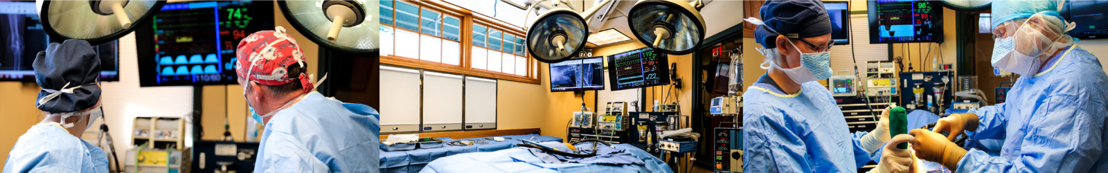
In patients who have failed to respond to conservative management or who are severely affected by hip dysplasia, surgery is typically necessary to achieve an acceptable level of comfort and mobility. A number of different surgeries can be performed to address hip dysplasia, and the best procedure for a specific patient will depend on their age, body size, extent of degenerative changes, and concurrent health conditions. Surgeries for hip dysplasia fall into two categories: those that address abnormalities present in affected puppies with the goal of preventing progression of hip dysplasia (Juvenile Pubic Symphysiodesis (JPS), Triple Pelvic Osteotomy (TPO), and Double Pelvic Osteotomy (DPO)), and those that aim to achieve a comfortable and functional limb in patients with advanced degenerative joint disease as a consequence of hip dysplasia (Femoral Head and Neck Ostectomy (FHO) and Total Hip Replacement (THR)).
Juvenile Pubic Symphysiodesis (JPS)
This surgery aims to induce a change in the orientation of the hip sockets as the immature pelvis grows in order to increase the surface area that is in contact with the weight-bearing portion of the femoral head. This change in the geometry of the hip joint decreases the abnormal forces that cause stretching, remodeling, and ultimately degneration of the hip joints over time. This is achieved by prematurely fusing the growth plate that runs along the midline of the bottom of the pelvis—called the pubic symphysis—using electrocautery. This prevents further growth in this location. As the rest of the pelvis continues to grow normally, the two sides of the pelvis are forced to rotate slightly downward, causing the hip sockets to assume a slightly more horizontal orientation. It is necessary to spay or neuter all patients undergoing this procedure due to its impact on a female’s ability to give birth as well as the high likelihood of passing on hip dysplasia to offspring.
This procedure will only be effective in young puppies who have yet to undergo signifcant growth of the pelvis. Typically, in puppies up to about 5 months old with signs of hip laxity, this surgery is effective in either preventing or significantly reducing the development of hip dysplasia as they grow. However, it is not possible to determine at such a young age which patients with hip joint laxity will go on to develop significant hip dysplasia without surgery, resulting in the possibility that some patients who have this surgery may not ultimately have needed it.
Triple Pelvic Osteotomy (TPO)
This surgery has the same goal as the Juvenile Pubic Symphysiodesis, to alter the orientation of the hip socket in order to improve its coverage of the femoral head. With the Triple Pelvic Osteotomy, this is achieved by making cuts through the pelvis in three places surrounding the hip socket. The resulting bone fragment containing the hip socket is then rotated slightly horizontally to achieve the desired orientation, and is then secured back to the rest of the pelvis using a specially-designed surgical plate and screws.
This procedure is only effective in dogs that do not yet have significant remodeling or any arthritic changes. Typically, only patients from 5 months old up to about 12 months old meet these criteria. This is a relatively invasive surgery, and has a moderate risk of complications. Similar to the JPS, it is important to spay or neuter any patient that undergoes this procedure. Additionally, there is a similar question with this surgery of whether many patients with joint laxity as puppies will really go on to develop symptomatic hip dysplasia, potentially resulting in some patients undergoing an unnecessary surgery. At this time, there is not a reliable way to predict the progression of symptomatic hip dysplasia in a puppy with signs of hip joint laxity.
Double Pelvic Osteotomy (DPO)
This surgery is very similar to the Triple Pelvic Osteotomy, but involves only two cuts through the pelvis instead of three. Sufficient rotation of the hip socket can be achieved despite the lack of the third cut. The DPO has the same limitations and requirements as the TPO, but does tend to have a lower rate of complications.
Femoral head and neck ostectomy (FHO)
This surgery involves removing the head and neck of the femur, so the hip joint no longer has a “ball.” The hip socket and pelvis are not altered. As the body heals, a fibrous “false joint” forms between the hip socket and the spot where the femoral neck was removed. With this procedure, early and consistent physical rehabilitation therapy is absolutely essential to a good functional outcome. Specifically, stretching and range-of-motion exercises are necessary to prevent hip flexor muscle contracture. If contracture does develop, it is usually permanent and severely limits limb use. For this reason, many patients are hospitalized for several days following their surgery to allow for frequent post-operative therapy sessions with one of our experienced physical rehabilitation technicians. When a patient is sent home, these exercises are demonstrated and detailed instructions are provided to ensure proper home care. With the correct post-operative therapy, the functional outcome of this procedure is generally excellent for small and medium-sized dogs, with full return to normal limb use, and can also be good for many larger breeds, especially if they are kept at a lean body mass.
Total hip replacement (THR)
Total Hip Replacement surgery involves the complete replacement of the ball and socket of the hip joint with metal and synthetic implants. The head and neck of the femur are removed, and a metal implant shaped like a healthy femoral head and neck is securely anchored into the femur. The hip socket’s joint surface is removed, and a prosthetic cup is implanted. This is a complex surgery that requires special expertise and careful surgical technique.
Total Hip Replacement surgery is an option for any fully-grown patient affected by hip dysplasia. Most patients experience significantly improved comfort and limb use rapidly after surgery, although physical activity must be limited until the bones and soft tissues around the hip have had sufficient time to heal. When performed by an experienced surgeon, the risk of complications is low, with joint luxation (dislocation), femur fracture, surgery site infection and implant loosening being the most common. Close adherence to post-operative instructions and periodic post-surgical check-ups will help to minimize the development of complications.
What is the prognosis for hip dysplasia in dogs?
Hip dysplasia often develops into a debilitating condition that severely impacts a dog’s quality of life. With appropriate treatment, however, a dog’s mobility and comfort can be dramatically improved, allowing them to live a full and comfortable life.
Let our highly trained and experienced team of veterinarians and veterinary technicians help you keep your puppy as happy and healthy as they can be.
Call the Animal Clinic of Billings and Animal Surgery Clinic to schedule your pet’s next wellness examination with one of our veterinarians today!
406-252-9499 REQUEST AN APPOINTMENT