Veterinary Ultrasound, X-Ray, MRI, CT Scans and more
What is veterinary special diagnostic imaging for dogs and cats?
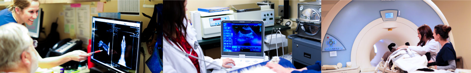
 Veterinary diagnostic imaging refers to Ultrasound, Radiography (X-rays), MRI, Doppler Echocardiogram, and CT Scans. Veterinary diagnostic imaging enables us to detect illnesses and abnormalities in dogs and cats that cannot otherwise be appreciated and to do so in a non-invasive and non-painful manner. Ultimately, the goal of any specialized form of diagnostic imaging is to definitively detect an abnormality and determine its extent in the body without resorting to more invasive interventions, such as surgery.
Some imaging modalities, such as MRI and CT scans, require sedation or anesthesia because a dog or cat must be completely still to allow for highly detailed images to be acquired. Our veterinarians are then able to evaluate these images to establish a diagnosis and determine the best course of treatment.
As part of our diagnostic services, The Animal Clinic of Billings and Animal Surgery Clinic is equipped with digital X-ray and digital X-ray monitoring for both general and dental X-rays. Our doctors also perform ultrasound examinations in in-house and routinely coordinate MRI and CT scans.
The Animal Clinic of Billings is also equipped with a cutting-edge diagnostic laboratory to provide real-time lab results. This allows us to evaluate a patient’s status and implement appropriate treatments quickly.
When are ultrasound, X-ray, and MRI necessary for dogs and cats?
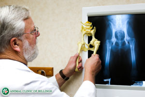
If the veterinarian suspects your dog or cat has an internal abnormality after performing a physical examination, diagnostic imaging is typically the next step to acquire more information. It’s important to understand that for some conditions, multiple types of diagnostic imaging procedures may be required to reach a definitive diagnosis. X-rays are typically the first type of diagnostic imaging performed. If a broken bone (fracture) is suspected, X-Rays are taken both of the injured limb as well as the opposite limb for comparison.
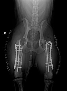
Your veterinarian will use the X-Rays to establish the best treatment plan. If surgery is necessary, the X-rays are used to determine the best strategy for internal stabilization, including implant type(s) and the size of plates, pins, or screws required. If X-rays are inconclusive or the problem resides in the soft tissue of the dog or cat, then an ultrasound or an MRI may be necessary to get a more thorough look at the area in question.
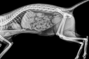 Let’s say you bring your dog or cat in because he or she is vomiting. One of our veterinarians will likely start by taking an X-ray to rule out the possibility of an intestinal obstruction due to the ingestion of a non-digestible foreign body. If the X-ray shows potential signs of intestinal obstruction but is not definitive, then either an ultrasound or a contrast X-ray study may be necessary to confirm the diagnosis before proceeding to surgery.
In a contrast GI X-ray study, the patient is given a solution containing barium to drink. Barium is bright white on x-rays, and through serial X-rays of the abdomen, the barium can be observed making its way through the stomach and intestines. If it stops abruptly at any point, this indicates the presence of a blockage.
Multi-modal imaging allows for greater confidence in a diagnosis and treatment plan, which is particularly important when the treatment is invasive or involves risk. Different diagnostic imaging modalities provide various types of information about an abnormality that can all be integrated to generate the most complete picture of what is going on.
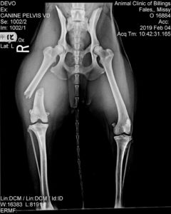 As another example, if your dog is not using his or her hind limb due to pain in their knee, possible causes include the following:
- Torn cruciate ligament
- Torn meniscus
- Torn collateral ligaments
- Luxating patella
- Patellar tendonitis
- Osteoarthritis
- Joint infection
- Immune-mediated joint disease
- Avulsion of the tibial tuberosity
X-rays of the dog’s knee often show signs of soft tissue swelling or fluid buildup in or around a specific area but may not pinpoint which structure is damaged. Therefore, in some cases, more advanced imaging with an MRI or CT scan may be necessary to make a definitive diagnosis.
Because an MRI is much more expensive than X-rays, we will almost always start with X-rays and resort to more expensive imaging modalities only if there is uncertainty about the diagnosis. In most cases, our veterinarians identify the problem from the initial X-rays and ultrasound and can confidently determine the best treatment protocol for your dog or cat.
How many types of diagnostic imaging procedures are there?
There are six types of veterinary diagnostic imaging procedures our veterinarians may utilize to assist in the diagnosis of your dog or cat’s condition. They include:
- X-ray
- Ultrasound
- MRI
- CT Scan
- Doppler Echocardiogram
- Endoscopy
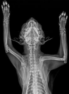
X-rays
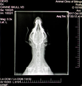
X-rays, also known as radiographs, have been used to diagnose medical problems in dogs, cats, humans, and many other animals for over 100 years. X-rays are the most commonly used form of diagnostic imaging in veterinary medicine because they are more affordable than different types of diagnostic imaging and they are beneficial for accurately diagnosing a wide variety of both bone and soft tissue abnormalities throughout the body.
 Today, digital radiography has become the gold standard for how radiographic images are obtained. Digital X-rays are beneficial for diagnosing and monitoring a wide variety of conditions. The Animal Clinic of Billings and Animal Surgery Clinic is equipped with both dental and general digital radiographic imaging.
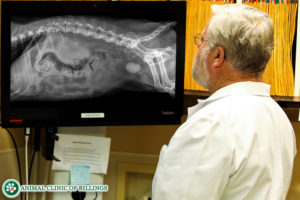
Modern digital X-rays expose the patient to significantly less radiation and eliminate the use of harsh processing chemicals. Our x-ray machines generate digital images that are stored immediately and permanently to your pet’s computerized medical record. The images can be enlarged, rotated, and otherwise manipulated to more easily visualize and diagnose skeletal and soft tissue diseases. Additionally, the images can be burned to a CD for you to take home, or sent to you via email.
X-rays are used to determine the presence of many internal conditions that cannot be otherwise detected, such as:
- Pulmonary diseases such as pneumonia and asthma
- Airway diseases such as bronchitis and collapsing trachea
- Heart disease and congestive heart failure
- Broken bones (fractures)
- Gastrointestinal foreign bodies
- Internal masses/tumors
- Joint diseases such as arthritis
- Kidney and bladder stones
- Jaw bone loss due to periodontal disease and tooth root abscesses
- and many more
Ultrasound on dogs and cats
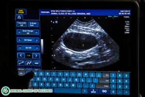
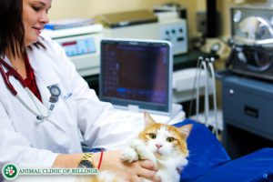 The second most commonly used type of veterinary diagnostic imaging is ultrasound. Ultrasound is a non-invasive, non-painful technique that produces a real-time image of your pet’s internal organs by emitting high-frequency sound waves through a sensor. Ultrasound helps to diagnosis abnormalities that don’t show up as well on X-rays, such as a mass hidden within an organ or the presence of thickened intestinal walls. Ultrasounds are typically not stressful for your dog or cat and take anywhere from 15 to 30 minutes to perform.
 Often times, ultrasound can be more accurate than x-rays because where x-rays show still images that detail the contours and density of hard structures in the body, ultrasound provides a movie of soft tissues and what’s happening inside your dog or cat’s body. This enables us to better assess things like heart problems and diseases of the abdominal organs, such as stomach and intestinal problems, liver and kidney disorders, disorders of the urinary bladder and reproductive system, adrenal disease, and evaluate the status of things like important glands and testicles.
Ultrasound is oftentimes also needed to use in conjunction with other diagnostic tools for comparative analysis. For example, if one of our veterinarians detects a lesion on your pet by first taking an x-ray, an ultrasound can then determine the origin of the lesion. In most cases, the ultrasound can then show us whether the lesion has spread to any other parts of your dog or cat’s body.
Ultrasound is also used to collect diagnostic specimens, including fluid and tissue samples. Using the ultrasound image as a guide, our veterinarians can perform surgical biopsies without the need for any major surgery, and oftentimes, your cat or dog is able to go home the same day. Ultrasound biopsies are non-painful and minimally invasive that have few complications compared to undergoing surgery for a biopsy. However, ultrasound biopsies provide only tiny samples and sometimes do not yield a definitive diagnosis; therefore, in some cases, a surgical biopsy is required.
What happens to a dog or cat during an ultrasound procedure?
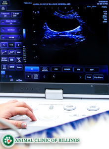
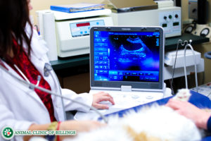
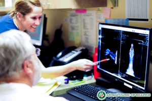
During an ultrasound, your dog or cat is carefully placed in a padded trough on a table. Your veterinarian then applies alcohol or ultrasound gel to the skin surface in the area of interest, and gently presses a small probe against the area. This probe emits digital sound waves to produce a diagnostic image of the underlying interior structures. The veterinarian moves the probe manually until a clear picture of the targeted internal area is displayed on the screen. The images generated by these sound waves are viewed in real-time on a computer screen, and can also be stored and saved in a computer for future viewing.
In modern scanning systems like those of the Animal Clinic of Billings and Animal Surgery Clinic, veterinarians can integrate the information from an ultrasound to determine a definitive diagnosis for your dog. They can then devise the most effective treatment protocol to achieve the best possible outcome.
CT Scans for dogs and cats
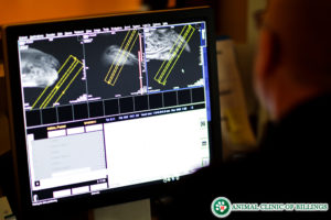
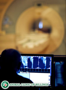
A CT scan uses technology similar to that of conventional X-rays, but much more advanced, to generate a three-dimensional view of the body. CT scans are particularly useful for imaging bones, joints, and the chest. A dog or cat must be sedated for a CT scan to ensure that they remain still for the duration of the scan. At the Animal Clinic of Billings, we routinely coordinate CT scans to diagnose a wide variety of injuries and illnesses.
Veterinary MRI for dogs and cats in Billings MT
 The Animal Clinic of Billings and Animal Surgery Clinic is proud to provide state-of-the-art veterinary MRI services in Billings for our canine and feline patients.
Because of the unique nature of MRI scanning and the prohibitive cost of the equipment, the Animal Clinic of Billings and Animal Surgery Clinic has established a partnership with a locally owned and operated Billings-based MRI facility designed for human care. This allows us to provide dogs and cats with the most cutting edge MRI technology available.
What is a dog or cat MRI?
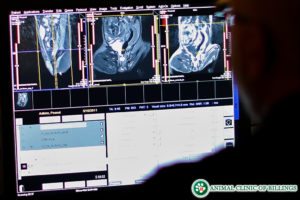
Magnetic Resonance Imaging (MRI) uses a powerful magnet–40,000 times stronger than the earth’s magnetic field—to generate detailed images of the body. While it is potent, an MRI does not emit radiation and is an extremely safe procedure.
How does a veterinarian perform a dog or cat MRI?
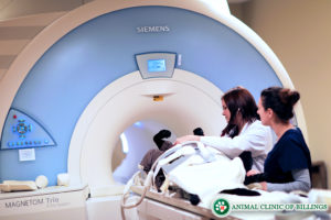
 During an MRI, the dog or cat is positioned to lie flat on a table that moves into the center of a domed-shaped MRI machine. After removing any metal from the patient (such as a collar), the vet will anesthetize the dog or cat and monitor him or her throughout the scan. Because it does take longer than an X-ray or CT scan to acquire the necessary images, heavy sedation or anesthesia is required to ensure a pet does not move, which would result in distorted and non-diagnostic images.
The MRI itself works by using a magnetic field and radio waves to create images. The procedure displays abnormalities, injuries, and diseases that may not be seen with any other method, with picture clarity and detail superior to other types of diagnostic scans.
An MRI typically takes more than an hour to complete, so the dog or cat is under anesthesia for a while, but rest assured that one of our veterinarians is always there to closely monitor the dog or cat’s condition and ensure everything goes as smoothly as possible.
What can a veterinarian see from a dog or cat MRI?
An MRI pinpoints individual differences in a dog’s tissue density, which can reveal tumors or other abnormalities that are difficult to see in ultrasound or CT scans. Sometimes, the patients are injected with a safe contrast material during an MRI to allow for even greater visual detail to highlight differentiating between structures or features within the dog or cat’s body.
MRI is commonly used to diagnose numerous conditions, including:
- Brain pathologies (trauma, tumors, malformations, strokes, and infections)
- Vertebral abnormalities
- Intervertebral disk disease (IVDD)
- Spinal cord injuries and tumors
- Bone, ligament, and tendon injuries and illnesses
- Joint diseases
- Growths or infections in the sinuses
- Vascular disease and blood clots
- Tumors, infections, injuries, and other disease processes within internal organs
Is an MRI safe for dogs and cats?
Unlike X-rays and CT scans, MRI scans do not emit radiation, so it’s considered to be safer than imaging systems. The MRI procedure also has no known negative side-effects on dogs, cats, or people. Regardless, we take every safety precaution to minimize the risk of complications to our MRI patients.
We check bloodwork before administering sedation or anesthesia to detect any conditions that might increase the risk of anesthesia, such as kidney or liver dysfunction. During the scan, we monitor our patients closely to ensure they are tolerating the medications. Rest assured that if an MRI is needed for your dog or cat, he or she is safe hands at the Animal Clinic of Billings and Animal Surgery Clinic.
Doppler echocardiograms on dogs and cats
 Another specialized diagnostic tool is a Doppler echocardiogram. Echocardiograms produce precise ultrasound images of the heart and cardiovascular system. An echocardiogram can show the contractions of the heart, the movement of its valves, and even the blood flowing through the arteries, all in real-time.
If your dog or cat is diagnosed with a heart murmur or some other heart-related problem, our veterinarians and staff will help you through every step to achieve the best possible outcome for your feline or canine companion. At the Animal Clinic of Billings and Animal Surgery Clinic, we coordinate echocardiograms to diagnose and monitor heart disease.
Endoscopy on dogs and cats
An endoscope is a small camera mounted on a fiber optic cable that veterinarians use to examine the GI tract visually. Using this tool, a veterinarian can directly visualize the interior of the esophagus, stomach, the first segment of the small intestine, and the colon.
Biopsies can be collected to diagnose GI diseases, and in some cases, foreign objects can be removed, enabling a patient to avoid needing surgery. The patient must be anesthetized before undergoing an endoscopic procedure. The endoscope is a handy tool that, in some cases, allows for the diagnosis and treatment of a patient without surgery.
 Let our highly trained and experienced team of veterinarians and veterinary technicians help you keep your pet as happy and healthy as they can be.
Call the Animal Clinic of Billings to schedule your pets next wellness examination!
406-252-9499 REQUEST AN APPOINTMENT