Patellar Luxation
What is patellar luxation?
Patellar luxation is a condition in which the patella (kneecap), which normally slides up and down within a groove running along the front of the femur within the knee joint as the knee is flexed and extended, pops out of its groove to one side. The kneecap can luxate either to the inside (medially) or the outside (laterally) of the groove. When the patella luxates, in addition to causing discomfort, it disrupts the normal function of the knee by pulling the quadriceps muscle group slightly to the side. This is a relatively common disorder in dogs, but is seen only rarely in cats.
What causes patellar luxation?
Patellar luxation can be caused either by genetic factors or by traumatic injury. In about 75% of cases in dogs, the patella luxates medially (to the inside). Most cases of medial patellar luxation are genetic in origin and occur in toy and small breed dogs. Affected individuals are born with anatomic abnormalities including coxa vara (where the femoral neck is too horizontally oriented) and decreased femoral neck angle of anteversion (where the angle between the long axis of the femoral neck and the transverse axis of the knee joint is too small, resulting in mild external rotation of the knee relative to the hip). These anatomic abnormalities cause inward (medial) displacement of the quadriceps muscle group and patella. As the limb is used, the abnormal forces generated by this displacement cause the leg bones to bow outward at the knee as the individual grows, which worsens the misalignment. In addition, the lack of the normal pressure of the patella gliding in the femoral trochlear groove results in underdevelopment of this important structure. Breeds at particularly high risk of medial patellar luxation include the Yorkshire terrier, Boston Terrier, Maltese, Papillon, Shih Tzu, Chihuahua, Pug, Pekingese, Miniature Pinscher, Japanese Chin, Manchester Terrier, Affenpinscher, English Toy Spaniel, and Brussels Griffon.
Lateral patellar luxation is also usually the result of genetic abnormalities, but primarily occurs in large and giant breed dogs. The anatomic abnormalities in lateral patellar luxation include coxa valga (where the femoral neck is too vertically oriented), increased femoral neck angle of anteversion (where the angle between the long axis of the femoral neck and the transverse axis of the knee joint is too large, resulting in mild internal rotation of the knee relative to the hip), and outward rotation of the tibia (shin bone). These anatomic abnormalities cause outward (lateral) displacement of the quadriceps muscle group and patella. As the limb is used, the abnormal forces generated by this displacement cause the leg bones to bow inward at the knee as the individual grows, which worsens the misalignment. In addition, the lack of the normal pressure of the patella gliding in the femoral trochlear groove results in underdevelopment of this important structure. Breeds at particularly high risk of lateral patellar luxation include the Great Dane, Newfoundland, Irish Wolfhound, and Saint Bernard.
Traumatic cases of medial and lateral patellar luxation are usually the result of a fracture of the femur (thigh bone) that heals in improper alignment, resulting in abnormal forces similar to those seen with genetic luxations.
What are the signs of patellar luxation?
The signs of medial patellar luxation vary depending on the severity of the luxation. In milder cases, dogs display intermittent lameness that often comes and goes relatively abruptly. They may suddenly hold up their limb with the knee fully extended for several strides, and then resume using it normally. It is also common for affected individuals to “bunny hop,” or move both hind limbs together, when running. In more severe cases, dogs usually have a persistent lameness, and may assume a bow-legged posture and a crouched gait. Usually signs first appear in puppyhood or young adulthood. Both knees are affected in about 50% of cases.
The signs of lateral patellar luxation are similar to those of medial patellar luxation, but these patients often exhibit a “knock-kneed” stance and external rotation of their feet. Usually signs first appear by 18 months of age, and most affected dogs have luxation in both of their knees.
How is patellar luxation diagnosed?
Patellar luxation is diagnosed on physical examination. The veterinarian palpates the kneecap as they flex and extend the knee, and applies pressure to it in particular positions to determine whether it can be moved into and out of the groove. Based on this examination, the grade of the luxation is determined:
- Grade 1 – The patella can be pushed out of the groove, but immediately pops back in on its own. This can be an incidental finding in a dog without any clinical signs.
- Grade 2 – The patella pops out on its own, but returns to the groove with flexion of the knee.
- Grade 3 – The patella is always out of the groove, but can be pushed back in. After being pushed into its groove, it immediately pops back out.
- Grade 4 – The patella is always out of the groove and cannot be pushed back in.
If there is any uncertainty about the diagnosis, which may be the case if there is a large amount of scar tissue, swelling, or other evidence of disease present in the knee, sedated x-rays are taken to help clarify the diagnosis.
How is patellar luxation treated?
The best treatment for a case of patellar luxation depends on the grade of luxation and the severity of clinical signs.
Grade one luxations that are found incidentally and do not cause clinical signs typically do not require treatment unless they proceed to cause symptoms in the future. Most grade one luxations that do cause clinical signs can be managed medically with anti-inflammatory medications, joint health supplements and nutraceuticals, and weight management. Commonly-used anti-inflammatory medications include carprofen (Rimadyl), deracoxib (Deramaxx), firocoxib (Previcox), meloxicam (Metacam), and grapiprant (Galliprant). Supplements and nutraceuticals that are often helpful include polysulfated glycosaminoglycans (Adequan), glucosamine, chondroitin, omega-3 fatty acids, methylsulfonomethane (MSM), avocado/soybean unsaponifiables, curcumin, green-lipped mussel extract, MicroLactin, and boswellia. The impact of weight reduction cannot be overstated. Maintaining a lean body mass can significantly reduce the odds of a luxation becoming worse over time.
Grade two luxations can often be managed conservatively in the same manner as grade one luxations. If a grade two luxation causes frequent or persistent symptoms, surgical intervention may be indicated.
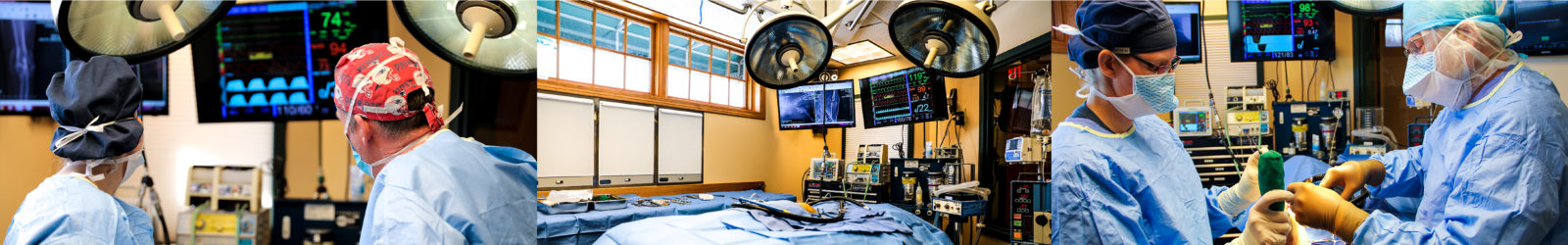
Surgery is the treatment of choice for all grade three and four luxations. The goal of surgical treatment is to correct the alignment of the quadriceps muscle group so the patella remains within its groove, restoring normal knee function.
Surgical treatment of medial patellar luxation involves a combination of several different surgical manipulations.
- A trochleoplasty is performed to increase the depth of the trochlear groove. This involves the removal of a wedge or block-shaped piece of cartilage and bone from the groove, followed by deepening of the resulting defect and replacement of the original fragment. Creating a deeper groove makes it more difficult for the patella to luxate.
- A lateral tibial tuberosity transposition is performed to shift the insertion point of the patellar tendon laterally. The patellar tendon extends from the end of the quadriceps musculature to the front of the upper tibia (shin bone), and houses the patella. Its attachment point on the tibia, called the tibial tuberosity, is the bony prominence palpable just below the knee joint. In a tibial tuberosity transposition, a cut (osteotomy) is made through the bone of the tibia to detach the tibial tuberosity. The fragment is then shifted slightly laterally (toward the outside) and secured in place with surgical pins. By moving the insertion point of the patellar tendon laterally, this procedure re-aligns the patella with its groove.
- A medial desmotomy, or vertical incision through the contracted connective tissues and joint capsule on the medial (inner) side of the knee joint, releases tension that has been helping to hold the quadriceps and patella in their medially displaced position.
- A lateral imbrication, in which the stretched portion of the joint capsule on the outer (lateral) side of the knee joint is tightened up, helps to further stabilize the patella.
Surgical treatment of lateral patellar luxation involves similar manipulations performed so as to achieve the opposite affects to those of the medial patellar luxation surgery.
- A trochleoplasty is performed in the same manner as for a medial patellar luxation.
- A medial tibial tuberosity transposition is performed to shift the insertion point of the patellar tendon medially.
- A lateral desmotomy is performed to release contracted soft tissues on the lateral aspect of the knee.
- A medial imbrication is performed to tighten up the stretched joint capsule on the medial aspect of the knee.
In some cases where the femur is significantly bowed, a bone-straightening surgery called a corrective osteotomy may be performed to improve the alignment of the quadriceps apparatus relative to the knee joint. This involves the removal of a precisely-measured wedge of bone, apposition of the cut ends, and plating of the bone to stabilize it while it heals in its new, straighter form. This is more frequently necessary with lateral luxations than medial luxations.
What is the recovery like after surgery?
Following surgery, physical activity must be restricted until the bones have fully healed. X-rays are taken at 4-6 weeks and again at 8-10 weeks to monitor the healing of the tibial tuberosity osteotomy. Most dogs are able to return to full activity after their 8-10 week recheck. Until then, only low-impact on-leash exercise is allowed in order to prevent excessive pull on the tibial tuberosity as it heals. For most dogs, this means two to three short, slow walks per day. You will also be instructed on how to perform range of motion exercises and stretches to prevent muscle contracture and joint stiffness during the healing process.
What are the potential complications of surgery?
The most frequently encountered complication is recurrence of the luxation. With medial patellar luxations, the risk of re-luxation is about 10% for grade three luxations and is greater than 30% for grade four luxations. Lateral patellar luxations are reported to recur around 20% of the time. Other potential complications include implant loosening or breakage, tibial tuberosity avulsion (pulling away from the rest of the tibia), movement of the trochlear wedge, rupture of the patellar ligament, fracture of the lateral femoral trochlear ridge, and progression of arthritis. Large breed dogs and obese dogs have a higher risk of complications than smaller breed and lean dogs.
What is the prognosis for patellar luxation?
The prognosis for dogs with mild patellar luxation that is managed conservatively is very good, although some degree of arthritis is likely to develop over time. With strict weight management and appropriate administration of medications, supplements, and nutraceuticals, the development of arthritis in these dogs is often minimal.
The prognosis for more severe patellar luxations that are treated with surgery is very good in most cases, with most dogs returning to normal or near normal limb function, although in a minority of cases a second surgery is necessary.
Let our highly trained and experienced team of veterinarians and veterinary technicians help you keep your cat as happy and healthy as they can be.
Call the Animal Clinic of Billings and Animal Surgery Clinic to schedule your pet cat’s next wellness examination with one of our veterinarians today!
406-252-9499 REQUEST AN APPOINTMENT
ANIMAL CLINIC OF BILLINGS AND ANIMAL SURGERY CLINIC
providing our region’s companion animals and their families what they need and deserve since 1981
1414 10th St. West, Billings MT 59102
406-252-9499