Intervertebral Disk Disease – IVDD
What is Intervertebral disc disease (IVDD)?
Intervertebral disc disease (IVDD) is a condition in which one of the discs separating two vertebrae pushes upward into the spinal canal, traumatizing and compressing the spinal cord and nerve roots. Intervertebral discs are made up of two parts: a core, called the nucleus pulposus, composed of a dense, gel-like matrix of water molecules and proteoglycans (water-loving molecules found in cartilage), and an outer ring of connective tissue (the annulus fibrosus). Intervertebral discs normally function to cushion and support the bones of the spinal column (vertebrae) as an individual flexes, twists, and/or compresses their neck and back in the course of daily activities.
The term Intervertebral Disc Disease actually encompasses four different but related disease processes: Intervertebral Disc Extrusion (also called Hansen Type I Disc Disease), Intervertebral Disc Protrusion (also called Hansen Type II Disc Disease), Acute Non-compressive Nucleus Pulposus Extrusion (ANNPE), and Hydrated Nucleus Pulposus Extrusion (HNPE).
Hansen Type I Disc Disease (disc extrusion) is caused by a degenerative process occurring within the core of the disc (the nucleus pulposus). An important component of this process is chondroid metaplasia, the ingrowth of cartilage-producing cells (chondrocytes) into the core of the disc. Over time, the core loses hydration, becomes brittle and eventually begins to mineralize. Along with these structural changes, the core loses its pressure-dissipating function, resulting in the application of increasingly uneven stresses to the surrounding annulus fibrosus. This causes microtears to accumulate in the annulus. Because the annulus is thinnest adjacent to the spinal canal, this is the portion of the annulus where a rupture will eventually occur, allowing the core material to be pushed upward into the spinal canal. While this is a gradual disease process, the rupture and extrusion of disc material happens suddenly, causing a contusion to the spinal cord as well as a variable degree of stationary cord compression.
Hansen Type II Disc Disease (disc protrusion) begins with the same type of degenerative changes to the core of the disc. In patients affected by this type of disc disease, however, the increased strain on the annulus causes it to thicken rather than rupture. As the annulus slowly thickens over time, it progressively protrudes into the spinal canal and eventually begins to compress the spinal cord.
Acute Non-compressive Nucleus Pulposus Extrusion (ANNPE) occurs when a small volume of normal healthy nucleus pulposus extrudes through a tear in the annulus fibrosus at a high velocity, causing a significant contusion to the spinal cord without any subsequent spinal cord compression. This type of disc disease is believed to be caused by excessive strain during vigorous activity.
Acute Compressive Nucleus Pulposus Extrusion (ACNPE), also called Hydrated Nucleus Pulposus Extrusion (HNPE), is quite similar to ANNPE, the primary difference being that a larger volume of nucleus pulposus extrudes, resulting in both spinal cord contusion and compression.
What causes Intervertebral Disc Disease?
The degenerative process (chondroid metaplasia) underlying Hansen Type I and Type II Disc Disease is believed to be genetic in origin, although in most cases, the genes involved have not been identified. ANNPE and HNPE, in which the disc itself is normal and healthy, are thought to be primarily the result of trauma during intense activity.
Who is at risk for Intervertebral Disc Disease?
Hansen Type I Disc Disease is encountered primarily in chondrodystrophic (short-legged) breeds, such as the Dachshund, Shih Tzu, Beagle, Pekinese, American Cocker Spaniel, Corgi, French Bulldog, and Lhasa Apso. In these breeds, disc extrusion most commonly occurs between three and six years of age, although it may be seen in any individual over about two years old. This type of disc disease also occurs occasionally in non-chondrodystrophic breeds, but in these individuals it tends to develop at an older age.
Hansen Type II Disc Disease most commonly affects older, large breed dogs, such as the German Shepherd Dog, Labrador Retriever, and Doberman.
Acute Compressive and Non-compressive Nucleus Pulposus Extrusions (ACNPE & ANNPE) are typically experienced by young active dogs during vigorous exercise.
What are the symptoms of Intervertebral Disc Disease?
The symptoms of Intervertebral Disc Disease vary with the type of disease, the location, and the severity of injury or compression to the spinal cord. Individuals affected by Hansen Type I (disc extrusion) along the back usually develop signs affecting the low back and hind limbs. These tend to have a rapid onset, usually hours to a few days. In mild cases, an individual may only have signs of back pain without any functional impairments. This pain may cause them to assume a hunched posture and to be less willing to engage in normal physical activity. More significant compression causes hind limb weakness, incoordination, and even paralysis, as well as loss of bladder and bowel control. In the most severe cases, an individual may lose the ability to feel pain in their hindquarters. Hansen Type I disc extrusions in the neck most commonly produce pain and muscle spasms that cause an individual to avoid turning or raising and lowering their head. They may yelp or whine apparently at random as a result of a slight movement triggering sudden pain. It is also not unusual to observe a limp in one or both of the front legs due to pain radiating down the shoulder and into the forelimb. In especially severe cases, weakness and incoordination in all four legs may be seen.
The symptoms of Hansen Type II (disc protrusion) are usually much more gradual in onset, developing over a period of months. Affected individuals typically demonstrate signs of discomfort, as well as progressive weakness and wobbliness in their back legs (if the affected disc is in their back) or in all four legs (if the affected disc is in their neck). This weakness may cause them to scuff their paws as they walk. They may also have difficulty rising from a sitting position and using stairs. Sometimes one side of the body is more affected than the other.
Acute Compressive and Non-compressive Nucleus Pulposus Extrusions (ACNPE & ANNPE) cause a sudden onset of symptoms, usually during exercise. It is not uncommon for affected individuals to suddenly yelp while running or playing, and abruptly display weakness or paralysis in one or more limbs. Signs are often more severe on one side of the body than the other. Unlike the other forms of disc disease, ACNPE and ANNPE do not typically cause signs of pain adjacent to the affected disc.
How is Intervertebral Disc Disease diagnosed?
Diagnosing Intervertebral Disc Disease starts with a complete physical and neurologic exam. Findings consistent with IVDD vary based on location and severity, and often include pain on palpation and manipulation of the neck or back, altered reflexes, weak or delayed responses to standard positioning tests, decreased or absent muscle tone, and decreased or absent pain sensation.
When exam findings suggest disc disease, the next step is to take sedated x-rays of the vertebral column. While intervertebral discs are not visible on x-rays, narrowing of the disc space and/or mineralization within the degenerating disc are present in many cases. These x-rays are also important to rule out other possible causes of spinal cord-related signs, such as a vertebral fracture or luxation, infection of a vertebra or disc, or a mass involving one of the vertebrae.
Once x-rays have been used to rule out a vertebral abnormality as the cause of symptoms, a definitive diagnosis is established with a CT scan or MRI. These advanced imaging modalities enable visualization of the spinal cord, intervertebral discs, and surrounding soft tissues. The location and degree of cord compression and the severity of spinal cord contusion can both be determined. In order to perform a CT scan or MRI, heavy sedation or general anesthesia is necessary as the patient must remain very still during image acquisition.
How is Intervertebral Disc Disease treated?
Treatment of Intervertebral Disc Disease depends on the type and severity of disease.
Hansen Type I IVDD (disc extrusion)
Hansen Type I IVDD (disc extrusion) in the neck is usually treated with conservative management if no neurologic deficits are present. This consists of strict confinement for four weeks to prevent further extrusion while the annulus fibrosus heals and to give the body a chance to break down some of the extruded material. Anti-inflammatory medication, pain medication, and a muscle relaxant are typically administered during this period. If signs have fully resolved after four weeks, physical activity is gradually reintroduced. Because of the risk of recurrence, a collar should never be used on an individual who has had a disc extrusion—a chest harness should be used instead. Additionally, dogs who have had a previous extrusion should not be allowed to jump onto or off of things for the rest of their lives, as this type of strain on the spine can precipitate another extrusion.
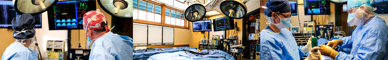
Patients with a disc extrusion in their neck who have neurologic deficits or who failed to respond to conservative management are best managed with surgery. The surgery that is most commonly performed to treat a cervical disc extrusion is called a ventral slot procedure. In this surgery, an incision is made along midline on the front of the neck. Tissues are retracted to either side to expose the affected disc and the vertebrae in front of and behind it. A rectangular window is carefully cut through the disc and a portion of each adjacent vertebral body. This window is deepened using a specialized drill until the spinal canal is accessed. The disc material can then be removed, relieving the compression. The window is left open, and the tissues of the neck are restored to their normal positions before closing the skin incision.
Hansen Type I IVDD (disc extrusion) in the back (thoracic or lumbar spine) is usually treated with conservative management if no neurologic deficits are present, or if only very mild deficits are present. Conservative management is the same as for cervical (neck) disc extrusion: strict confinement for four weeks to prevent further extrusion while the annulus fibrosus heals and to give the body a chance to break down some of the extruded material. Anti-inflammatory medication, pain medication, and a muscle relaxant are administered to aid in recovery. If signs resolve with conservative management, individuals can gradually return to full activity, but jumping and stairs should always be avoided in the future as these activities may precipitate another disc extrusion.
Patients with a disc extrusion in their back (thoracic or lumbar spine) who have moderate to severe neurologic deficits, as well as those that have failed to respond to conservative management, should be treated surgically. The surgery that is most commonly performed to address a thoracic or lumbar disc extrusion is called a hemilaminectomy. In this procedure, an incision is made along that back over the affected disc space. The muscles of the back are retracted away from the vertebrae on the side containing the majority of the extruded disc material. A window is cut into the side of the vertebrae in front of and behind the disc space, exposing the spinal canal. The extruded disc material is then carefully removed, relieving the spinal cord compression. A fat graft is placed in the window to protect the spinal cord and prevent scar tissue adherence. The muscles are returned to their normal positions, and the skin incision is closed.
Hansen Type II IVDD (disc protrusion)
Hansen Type II IVDD (disc protrusion) in the neck or back is generally treated conservatively if only pain and/or minimal neurologic deficits are present. Conservative management consists of a six to eight week period of activity restriction with medications to relieve pain and inflammation. A weight loss diet may also be implemented for overweight patients. Response rates to this type of treatment are generally good, although there is a high risk of recurrence of symptoms in the future.
Hansen Type II IVDD (disc protrusion) in the neck or back that causes moderate to severe neurologic deficits, or that has failed to respond to conservative management, may be treated surgically. In the neck, a ventral slot procedure is typically performed, while in the back, a hemilaminectomy is more common. In some cases of disc protrusion, the surgeon may determine that distraction and stabilization of the disc space is necessary, and this can be performed at the same time as decompression. This involves the use of screws, pins, wire, and/or bone cement to rigidly stabilize the two vertebrae adjacent to the affected disc space in a neutral position.
Acute Compressive Nucleus Pulposus Extrusion (ACNPE)
Acute Compressive Nucleus Pulposus Extrusion (ACNPE), also know as Hydrated Nucleus Pulposus Extrusion (HNPE), is treated in the same way as Hansen Type I disc extrusions, with conservative management for mildly affected patients and surgery for those more severely affected.
Acute Non-compressive Nucleus Pulposus Extrusion (ANNPE)
Acute Non-compressive Nucleus Pulposus Extrusion (ANNPE) is always treated medically rather than surgically. This is because there is no spinal cord compression to relieve; all of the clinical signs are caused by spinal cord contusion. Thus, regardless of the severity of the neurologic deficits, all cases are treated with rest, nursing care, and medications to reduce pain and inflammation until the spinal cord has a chance to heal, which may take anywhere from two to six weeks.
What post-operative care is required following surgery for Intervertebral Disc Disease?
Whether a patient has spinal surgery on their neck or their back, proper post-operative care is essential for a functional outcome. Following spinal surgery, all patients are hospitalized on IV fluids to maintain hydration and injectable medications for pain and inflammation. They are kept on thick bedding, and if unable to stand up and move around on their own, they are repositioned frequently to prevent pressure sores and lung compression. If bladder control is absent, a urinary catheter or gentle manual pressure is used to empty their bladder on a regular schedule. Patients are bathed as frequently as necessary to keep them clean and prevent skin irritation from developing. Once a patient is eating and drinking on their own and is comfortable on oral medications, they can be sent home. This may take as little as 24 hours, or as much as a week. Disc-related pain is typically significantly improved within 24 hours of surgery, and neurologic deficits often begin to improve within three to five days. Maximal recovery of nerve function can take anywhere from a couple of weeks to several months, and generally relates to the duration of symptoms before surgery.
At home, patients who are able to walk must be confined to strictly limit their physical activity while they heal. Only short, slow leash walks (with a chest harness rather than a collar) are permitted for the first few weeks after surgery, and they must be prevented from jumping, playing, or stair use. Slick surfaces should also be avoided. Raised food and water bowls should be provided to pets who have had neck surgery, so they do not need to stretch their neck down to the floor to eat and drink.
Patients who are not able to get up and move about on their own, as well as those that do not have complete bladder or bowel control, require more intensive care at home. This includes deep bedding that is kept clean and dry at all times, frequent repositioning to avoid pressure sores, and the use of sling support when taking them outside to use the bathroom. If a patient is not able to empty their bladder on their own, the owner is taught how to catheterize or manually express their bladder. Patients who are incontinent will need to be checked frequently for any urine or feces that might be on their hind end, and cleaned with warm water and a gentle cleanser whenever soiling is present.
The first follow-up appointment is typically scheduled for two to three weeks after surgery. At this appointment, the progress of healing and restoration of function will be assessed, and the stitches will be removed. Based on the patient’s progress, the veterinarian will determine if an increased level of physical activity can be introduced. For patients who have not yet recovered normal nerve function, a physical rehabilitation program will be implemented to facilitate neurologic recovery. Further recheck appointments are scheduled once a month until maximal neurologic improvement is achieved.
What are the potential risks and complications of surgery for Intervertebral Disc Disease?
Several complications can occur during or following surgery for Intervertebral Disc Disease. Because of the close proximity of the spinal cord and spinal nerves to the tissues that must be manipulated during surgery, there is a risk of temporary or permanent damage to these structures. If the large blood vessels located within the spinal canal are disrupted during surgery, significant hemorrhage may result. While this hemorrhage is rarely life-threatening, it often necessitates halting the procedure, closing the surgical site, waking the patient up, and waiting a day or two before attempting a second surgery to finish the procedure. Magnification, specialized instruments, and very careful and precise surgical technique help to minimize these risks.
In the immediate post-operative period, in addition to the potential for a surgical site infection, patients with significant neurologic deficits are at increased risk of developing pneumonia, urinary tract infections, and skin infections. These risks are minimized by providing intensive nursing care to patients who are immobile and/or incontinent.
What is the prognosis for Intervertebral Disc Disease?
The prognosis for a patient with Intervertebral Disc Disease depends on the type of disease as well as the severity of the neurologic deficits at the time of diagnosis.
Type I IVDD (disc extrusion) causing only pain and mild neurologic deficits has about a 75% chance of resolving with conservative management. Those cases that fail conservative management have a greater than 95% chance of resolution with surgery. Patients with Type I IVDD that is causing significant neurologic dysfunction (inability to walk, incontinence) but who are able to feel their affected limbs, have a 90% chance of returning to normal or near-normal function following surgery. Individuals who are unable to feel pain in their affected limbs have a much more guarded prognosis, with less than a 50% chance of recovery with surgery. This worsens to less than 10% if pain sensation has been absent for more than 24 hours. In a small fraction of the most severely affected individuals, a process called spinal cord myelomalacia occurs, resulting in the death of nerve cells in the spinal cord at the site of the initial contusion. This process is irreversible, and may progress up and down the spinal cord. Patients diagnosed with this condition have no hope of recovery, and are at a high risk of dying from respiratory paralysis; for this reason, euthanasia is recommended in cases of myelomalacia. It is also important to keep in mind that an individual who has experienced a disc extrusion is at an elevated risk of having another disc extrusion in the future. Avoiding jumping, stairs, and vigorous play indefinitely can delay the extrusion of other degenerating discs.
Patients with Type II IVDD (disc protrusion) causing pain and mild neurologic deficits have a 50% chance of resolution with conservative management. About half of patients who respond to conservative management will eventually relapse, and often respond favorably to another course of conservative treatment. Those with moderate to severe neurologic deficits rarely respond to conservative management, and unfortunately only 22% of patients will be restored to normal or near-normal function with surgery. Many patients experience partial improvement and can have a good quality of life with special support devices to aid in mobility.
Patients diagnosed with Acute Non-compressive Nucleus Pulposus Extrusion (ANNPE) have a very good prognosis for recovery of normal or near-normal function with conservative management. The risk of recurrence of this type of disc disease has not yet been well-defined.
Patients with Acute Compressive Nucleus Pulposus Extrusion (ACNPE; a.k.a. Hydrated Nucleus Pulposus Extrusion, HNPE) have similar prognoses to patients with Type I disc extrusions with the same degree of neurologic deficits. Like ANNPE, the risk of recurrence is unknown at this time.
Let our highly trained and experienced team of veterinarians and veterinary technicians help you keep your cat as happy and healthy as they can be.
Call the Animal Clinic of Billings and Animal Surgery Clinic to schedule your pet cat’s next wellness examination with one of our veterinarians today!
406-252-9499 REQUEST AN APPOINTMENT
ANIMAL CLINIC OF BILLINGS AND ANIMAL SURGERY CLINIC
providing our region’s companion animals and their families what they need and deserve since 1981
1414 10th St. West, Billings MT 59102
406-252-9499