
Canine Cardiology
 Do dogs get heart disease?
While dogs and cats do not develop the atherosclerosis and consequent heart attacks so commonly encountered in humans, they are susceptible to a variety of other types of heart disease. These can be broadly classified into problems with the heart valves, the heart muscle itself, and the electrical wiring of the heart.
What are the symptoms of heart disease in dogs?
The most common symptoms of heart disease in dogs and cats include coughing, exercise intolerance (getting tired easily), breathing rapidly when inactive, and fainting episodes. The development of any of these signs should prompt a veterinary visit for an exam and any necessary testing.
Heart Valve Abnormalities
-
What Causes Heart Valve Diseases?
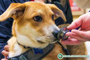
Abnormalities of the heart valves can be congenital (from abnormal development before birth), infectious, or degenerative (common in senior toy and small breed dogs). Normal heart valves function to prevent backward flow of blood as the heart pumps, ensuring that all of the blood delivered to the heart is propelled in the right direction each time the heart muscle squeezes and relaxes. Valve diseases result in backward leakage of blood through one or more valves. Over time, this leakage causes the heart to become overloaded as it has to pump harder and faster to keep enough blood flowing in the correct direction. The backflow of blood through a leaking valve can often be heard as a murmur when a doctor listens to a patient’s heartbeat with a stethoscope.
-
What are the Symptoms of Heart Valve Disease?
Symptoms of heart valve disease most often include a persistent soft cough (often worst in the morning), exercise intolerance, and increased respiratory rate and effort.
-
How are Heart Valve Diseases Diagnosed?
The diagnosis of a heart valve disease begins with a physical exam. The volume, location, and timing of a heart murmur can narrow down the list of possible causes. In order to definitively diagnose one of these diseases and determine severity, an echocardiogram (ultrasound of the heart) is performed. This allows for direct observation of valve movement and blood flow, as well as measurements of the heart chambers and strength of contractions. Chest X-rays, which reveal the size and external contours of the heart, can also provide information about which valve is involved based on which portion of the heart is enlarged, but do not provide as much information as an echocardiogram.
-
 How are Heart Valve Diseases Treated?
 How are Heart Valve Diseases Treated?
Treatment depends on the cause of the valve disease. Some types of congenital valve abnormalities can be corrected with cardiac catheterization procedures (such as balloon valvuloplasty), available at some specialty centers. Antibiotics are essential for the treatment of infectious endocarditis, and long-term heart medications may be necessary if irreversible damage has occurred. Degenerative valve disease, the most common type of valve disease encountered in dogs, while not curable, can be treated medically for extended periods of time with the goal of decreasing the strain on the heart while maintaining sufficient blood flow.
Diseases of the Heart Muscle
-
What Causes Heart Muscle Diseases in Dogs?
Diseases of the heart muscle include congenital abnormalities such as atrial and ventricular septal defects (holes between two chambers of the heart), as well as heart muscle diseases that develop at some point in a dog or cat’s life, such as dilated cardiomyopathy (DCM), hypertrophic cardiomyopathy (HCM), and arrhythmogenic right ventricular cardiomyopathy (ARVC).
-
Congenital Structural Abnormalities
Often structural defects present at birth, such as septal defects, are detected on a puppy’s first pediatric examination. These abnormalities cause heart murmurs that can be heard with a stethoscope. An echocardiogram can then be performed to determine the size and location of the defect, and to determine the best treatment (if any is necessary) and prognosis.
Dilated Cardiomyopathy (DCM)
-
What is Dilated Cardiomyopathy?
Dilated cardiomyopathy is a disease that causes abnormal stretching out of the heart walls, causing the heart to become distended and weak, so it is not able to pump as much blood with each heartbeat. Ultimately, the heart becomes unable to keep up and blood backs up into the lungs, causing congestive heart failure. The stretching of the heart chambers can also result in the development of an abnormal heart rhythm, leading to serious complications.
-
What Causes Dilated Cardiomyopathy?
Usually this disease is caused by a genetic predisposition found in specific large and giant breed dogs (Doberman Pinscher, Great Dane, Irish Wolfhound, Newfoundland, Giant Schnauzer, etc.), although a dietary deficiency in the amino acid taurine can also cause DCM. Recently this disease has also been linked to feeding grain-free diets, although the nature of this link is still being researched.
-
What are the symptoms of Dilated Cardiomyopathy?
The most common symptoms of DCM include exercise intolerance, weakness, increased respiratory rate and effort, and coughing. Fainting episodes (syncope), decreased appetite, and weight loss may also occur.
-
How is Dilated Cardiomyopathy Diagnosed?
Along with a complete physical exam, DCM is diagnosed using x-rays and an echocardiogram. An electrocardiogram (ECG) is also performed to look for abnormalities in the electrical activity of the heart that can accompany this disease.
-
How is Dilated Cardiomyopathy Treated?
While DCM is not curable, progression can be slowed with medications to decrease the load on the heart, increase heart muscle strength, and suppress arrhythmias. Early detection significantly improves prognosis.
Hypertrophic Cardiomyopathy (HCM)
-
What is Hypertrophic Cardiomyopathy?
 What is Hypertrophic Cardiomyopathy?
Hypertrophic Cardiomyopathy is a disease that causes a thickening of the walls of the left ventricle of the heart (the main chamber pumping oxygenated blood out to the body), which decreases the volume of blood than can enter and leave the heart with each beat.
Backup of blood behind the left ventricle causes dilation of the upstream chamber (the left atrium) and stretching of the valve between these two chambers. Backflow through this valve exacerbates the heart’s difficulty in delivering sufficient blood to the body and predisposes the patient to the formation of blood clots, which can lodge in blood vessels throughout the body, a potentially fatal complication.
-
What Causes Hypertrophic Cardiomyopathy?
HCM, which is seen primarily in cats, has a genetic basis in the Maine Coon and Ragdoll, although it is seen in many domestic shorthair/mixed breed cats as well.
-
What are the Symptoms of Hypertrophic Cardiomyopathy?
Symptoms of HCM can be very subtle, and include an increase in resting respiratory rate and effort and a decreased activity level. If a blood clot detaches from the heart, sudden loss of function in one or both hindlimbs may occur.
-
How is Hypertrophic Cardiomyopathy Diagnosed?
On physical exam, the presence of HCM is suggested by the presence of a murmur, although a significant percentage of affected cats do not have a murmur. Muffled lung sounds may indicate the buildup of fluid around the lungs. The definitive diagnosis of HCM requires an echocardiogram.
-
How is Hypertrophic Cardiomyopathy Treated?
Like DCM, HCM is not curable, but can be medically managed, in some cases for an extended period of time. Medications to improve cardiac relaxation, decrease the load on the heart, and thin the blood (to inhibit clot formation) slow the progression of disease and decrease the risk of secondary complications. Prognosis is best in cases that are detected early in the course of disease.
Arrhythmogenic Right Ventricular Cardiomyopathy (ARVC)
-
What is Arrhythmogenic Right Ventricular Cardiomyopathy?
Arrhythmogenic Right Ventricular Cardiomyopathy (ARVC), also known as “Boxer Cardiomyopathy,” is a condition in which the normal muscle forming the walls of one chamber of the heart (the right ventricle) is replaced over time with stiff and non-functional tissue, impairing its normal pumping function and causing dangerous arrhythmias.
-
What Causes Arrhythmogenic Right Ventricular Cardiomyopathy?
This disease has a genetic basis, and is most commonly diagnosed in Boxers, although it can be seen in other breeds, including the English Bulldog and Weimaraner.
-
What are the Symptoms of Arrhythmogenic Right Ventricular Cardiomyopathy?
The most common symptoms of ARVC are exercise intolerance and fainting spells.
-
How is Arrhythmogenic Right Ventricular Cardiomyopathy Diagnosed?
Because the arrhythmias that develop in patients with ARVC tend to come and go, it can be a challenge to definitively diagnose this condition. A physical exam and echocardiogram may not reveal any abnormalities, but are important for ruling out other kinds of heart disease. Usually, diagnosis of ARVC requires a 24-hour EKG (using a device called a Holter Monitor), which is able to record the episodes of abnormal rhythm that occur spontaneously throughout the day.
-
How is Arrhythmogenic Right Ventricular Cardiomyopathy Treated?
Treatment of ARVC is focused on normalizing the heart rhythm with anti-arrhythmic medications, and long-term survival is possible when good control of arrhythmias is achieved.
Cardiac Arrhythmia
-
What causes cardiac arrhythmia in dogs?
Many different diseases are defined by abnormal electrical activity within the heart. Arrhythmias such as atrial fibrillation/flutter, ventricular tachycardia, and ventricular fibrillation, often result from one of the previously mentioned disease processes. Others, such as atrial standstill, sick sinus syndrome, supraventricular tachycardia, AV block, and bundle branch blocks, may occur in the absence of any other heart abnormalities.
-
What are the Symptoms of a Cardiac Arrhythmia?
The most common symptoms of a clinically significant arrhythmia are exercise intolerance and collapse/fainting.
-
How is a Cardiac Arrhythmia Diagnosed?
Arrhythmias are diagnosed using an ECG; sometimes a short ECG lasting only a few minutes is sufficient to make a diagnosis, while in other cases a 24-hour-long ECG (called a Holter Monitor study) is necessary.
-
How is Cardiac Arrhythmia Treated?
Management of most of these conditions involves either medications to help normalize the heart rhythm or implantation of a cardiac pacemaker.

Let our highly trained and experienced team of veterinarians and veterinary technicians help you keep your dog as happy and healthy as they can be.
Call and schedule an appointment for your four-legged friend today.