Tibial Tuberosity Advancement (TTA)
What is a Tibial Tuberosity Advancement (TTA)?
A TTA is a surgical procedure performed to address a torn cranial cruciate ligament (CCL or CrCL). In order to understand this procedure, a review of canine stifle (knee) anatomy is in order. The stifle is composed of the bottom of the femur (thigh bone), which is shaped into two rounded condyles with a deep furrow running between them (the intercondylar fossa), the top of the tibia (shin bone), which provides a relatively flat surface with a mild downward slope from front to back (called the tibial plateau), and the patella (kneecap). As the knee flexes and extends, the femoral condyles rock back and forth atop the tibial plateau, and the patella slides up and down within the trochlear groove running along the front of the bottom of the femur. A number of ligaments are present within and around the knee joint to maintain stability as the limb is used. Two of these, the cranial and caudal cruciate ligaments, run between the intercondylar fossa of the femur and the middle of the tibial plateau within the joint. The cranial cruciate ligament (analogous to the anterior cruciate ligament, or ACL, in humans) starts far back on the femur and runs toward the front of the tibia, while the caudal cruciate does the opposite, starting toward the front of the femur and running to the back of the tibia. The cranial cruciate ligament performs the important job of preventing backward sliding of the femur on the sloped tibial plateau when the animal bears weight on the limb.
When the cranial cruciate ligament is torn, the stifle becomes unstable, as the femoral condyles slide backward on the tibial plateau whenever weight is placed on the affected limb. This causes pain and inflammation that, if not addressed, will ultimately result in significant degenerative joint disease. The TTA surgery aims to stabilize the knee by moving the tibial tuberosity, with its patellar tendon attachment, forward, so that the patellar tendon becomes perpendicular to the tibial plateau. The increased forward pull of the patella tendon when weight is placed on the affected limb helps to counteract the back-sliding force on the end of the femur that occurs when the cranial cruciate ligament is ruptured.
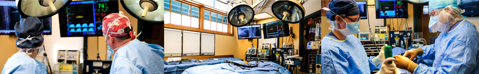
First, the surgeon enters the stifle joint and removes the damaged remnants of the cranial cruciate ligament. They also inspect the medial meniscus, which can become damaged as a result of joint instability. If damage is found, the affected portion of the medial meniscus is removed, leaving only healthy and functional meniscal tissue to cushion the knee. Next, the surgeon makes a straight vertical cut through the front of the tibia from the inner (medial) to the outer (lateral) surface, separating the tibial tuberosity from the rest of the tibia, leaving only the periosteum (thin flexible outer lining of the bone) at the bottom of the cut attached. This attachment serves as a hinge, and a small metal cage is inserted higher up in the cut to maintain a precisely-sized wedge-shaped gap between the two cut surfaces of the tibia. A special surgical plate is then secured across the gap to both bone segments, fixing them in their new configuration. Finally, a bone marrow graft or synthetic bone graft is placed into the gap to help facilitate bone ingrowth.
What patients can benefit from a TTA?
Almost any dog that has a torn cranial cruciate ligament can benefit from TTA surgery. While several different surgeries exist to address CCL tears, the TTA has shown to have results equal to or better than these other procedures. In particular, the TTA and TPLO are superior to other options for large and giant breed dogs, who may put too much strain on other stabilization measures. The best surgical option for a specific patient will depend on a number of variables, including age, breed, body mass, activity level, and the patient’s specific stifle anatomy. By evaluating these factors, the surgeon will be able to advise you on the best surgery for your pet.
What post-operative care is required?
Because the tibia bone requires eight to twelve weeks to fully heal in its new configuration, the post-operative recovery period is relatively prolonged. In the absence of complications or concurrent orthopedic or neurologic problems, a patient can be permitted to return to full activity on average three to four months after surgery. X-rays are taken at four to six weeks and again at eight to ten weeks after surgery to monitor the healing of the osteotomy.
During the first month after surgery, only short, slow leash walks are allowed. It is essential to avoid any running, jumping, and playing with other dogs during this period, as repeated excessive pull on the tibial tuberosity by the quadriceps apparatus can result in plate breakage or loosening, with subsequent displacement of the tibial tuberosity out of its desired position. This is a potentially serious complication that would require additional surgery to address. During this period, physical therapy exercises such as range of motion and stretches will be utilized to prevent joint stiffness and muscle contracture. After the first month, the length of leash walks can be gradually increased, and active physical rehabilitation exercises will be added to start building up muscle mass and encourage controlled weight bearing. Only after x-ray confirmation of advanced bone healing can patients be permitted to exercise off-leash, starting with only brief sessions and gradually increasing the duration and intensity until they are back to full activity.
What are the potential risks and complications of the TTA?
The overall complication rate for the TTA is comparable to that of the TPLO, at about 15%. Potential complications include surgical site infection, patellar luxation, loosening or breakage of the surgical implants, and tibial fracture. Most complications can be successfully resolved with medications and physical rehabilitation therapy, but in a small number of cases, a second surgery may become necessary.
What is the prognosis with this surgery?
With appropriate post-surgical management throughout the first few months after surgery, the prognosis is very good for return to normal or near-normal function. The outcome is generally comparable to that of the TPLO surgery. It is important to understand that degenerative changes that have already developed in the joint cannot be reversed, and some degree of arthritis will develop even with surgical stabilization. This, however, is much less severe than it would be without surgery, and is typically successfully managed with weight management, joint supplements, nutraceuticals, and anti-inflammatory medication as needed.
While a dog who has had a TTA has a lower risk of going on to develop a meniscal tear than a dog who has not had surgical stabilization, there is still about a 10% chance of medial meniscal injury in the future. This usually presents as a sudden-onset lameness developing up to a year after surgery, and may necessitate another procedure to remove the damaged portion of the meniscus. This surgery is much simpler than the TTA and has a much more rapid recovery.
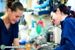
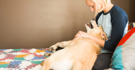
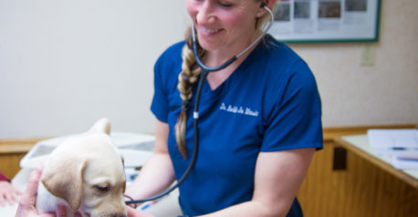
Let our highly trained and experienced team of veterinarians and veterinary technicians help you keep your cat as happy and healthy as they can be.
Call the Animal Clinic of Billings and Animal Surgery Clinic to schedule your pet cat’s next wellness examination with one of our veterinarians today!
406-252-9499 REQUEST AN APPOINTMENT
ANIMAL CLINIC OF BILLINGS AND ANIMAL SURGERY CLINIC
providing our region’s companion animals and their families what they need and deserve since 1981
1414 10th St. West, Billings MT 59102
406-252-9499